Pathologic Fracture in a Subchondral Cystic Lesion at the Superior Acetabulum
- Affiliations
-
- 1Department of Orthopedic Surgery, Seoul National University College of Medicine, Seoul, Korea.
- 2Department of Orthopedic Surgery, College of Medicine, Jeju National University, Jeju, Korea. kanguking@yahoo.co.kr
- KMID: 2185586
- DOI: http://doi.org/10.4055/jkoa.2010.45.2.160
Abstract
- Subchondral cyst is a benign cystic lesion, and it is often found in patients with rheumatoid arthritis or osteoarthritis. However, its occurrence in those joints without preexisted disease is rare. Furthermore, there has been no report in the medical literature regarding pathologic fracture in the bony plate of a subchondral cyst at the superior acetabulum. We report here on a case of the 44-year-old woman for whom a pathologic fracture was found in the bony plate of a subchondral cyst at the superior acetabulum.
MeSH Terms
Figure
-
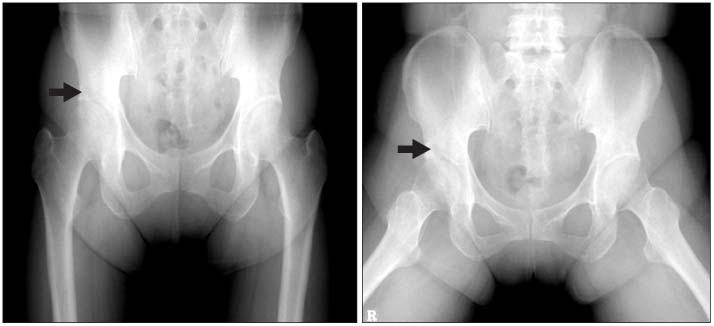
Figure 1 Both hip anteroposterior and frog leg radiographs obtained two months after the onset of pain in the right hip show ill-defined osteolytic lesion with focal poorly delineated right acetabular cortex.
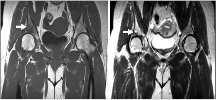
Figure 2 Coronal T1 and T2-weighted magnetic resonance images obtained at about two weeks later show that subchondral bone plate of right superior acetabulum is destructed and heterogeneous low signal intensity on T1-weighted image, which converted to heterogeneous high signal intensity at T2-weighted image. There are diffuse bone marrow signal change and enhancement at superior acetabulum and ilium.
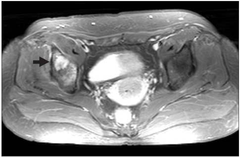
Figure 3 Axial T2-weighted enhanced magnetic resonance image obtained at about two weeks later shows diffuse bone marrow signal change at ilium and ill-defined round enhancement of the anterolateral aspect at right superior acetabulum.
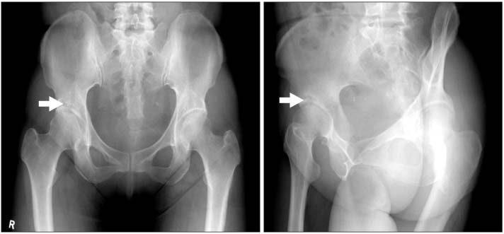
Figure 4 Pelvis anteroposterior and oblique radiographs obtained twenty seven months after the onset of pain in the right hip show residual cortical irregularity of right acetabulum.
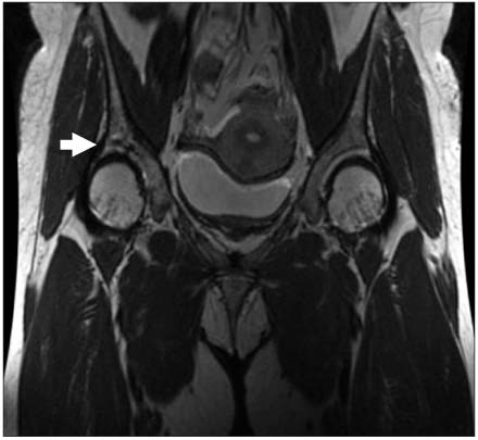
Figure 5 Coronal T2-weighted fat suppression magnetic resonance image obtained twenty seven months after onset of pain in the right hip does not show reactive change such as bone marrow edema and synovial enhancement. Also T2-weighted magnetic resonance image shows that destructed bony plate of superior acetabulum is migrated.
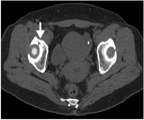
Figure 6 Axial computed tomographic image obtained twenty seven months after onset of pain in the right hip shows a subchondral cyst with subchondral bone plate fracture at right superior acetabulum and shows medullary sclerosis around subchondral cystic lesion.
Reference
-
1. Woods CG. Subchondral bone cysts. J Bone Joint Surg Br. 1961. 43:758–766.
Article2. Schajowicz F, Sainz MC, Slullitel JA. Juxta-articular bone cysts (Intra-osseous ganglia):a clinicopathological study of eighty-eight cases. J Bone Joint Surg Br. 1979. 61:107–116.3. Eggers GWN, Evans EB, Blumel J, Nowlin DH, Butler JK. Cystic change in the iliac acetabulum. J Bone Joint Surg Am. 1963. 45:669–722.
Article4. Motomura G, Yamamoto T, Miyanishi K, Shirasawa K, Noguchi Y, Iwamoto Y. Subchondral insufficiency fracture of the femoral head and acetabulum: a case report. J Bone Joint Surg Am. 2002. 84-A:1205–1209.5. Silas SI, Resnik CS, Levine AM. Bilateral primary cystic arthrosis of the acetabulum. A case report. J Bone Joint Surg Am. 1996. 78:775–778.
Article6. Resnick D. Diagnosis of bone and joint disorders. 1995. 3rd ed. Philadelphia: WB Saunders;3878–3882.
- Full Text Links
-
- Actions
-
Cited
- CITED
-
- Close
- Share
-
- Similar articles
-
- Sbuchondral Curettage & Bone Peg Fixation in Osteochondral Fracture of The Talus
- Reduction Technique of Dome Impaction Using the Modified Stoppa Approach: A Technical Note
- Treatment of bone cystic change with femoral head fracture in Neurofibromatosis patient
- Medialization of Cementless Acetabular Components after the Removal of Subchondral Bone in Total Hip Arthroplasty
- Stress fractures in calcaneus and juxtatectal region of the acetabulum : case report
