Osteosarcoma of the Metacarpal Bone
- Affiliations
-
- 1Department of Orthopaedic Surgery, Pusan National University School of Medicine, Busan, Korea. osteokim@yahoo.co.kr
- KMID: 2185254
- DOI: http://doi.org/10.4055/jkoa.2014.49.1.58
Abstract
- Osteosarcoma commonly develops around the knee joint, and rarely in the hand. Patients with osteosarcoma of the hand often present with pain and swelling, and osteosarcoma of the hand has a biological behavior that differs from that of osteosarcoma at conventional sites. However, although it usually occurs in the older age group, compared with conventional osteosarcoma, the most common sites of hand osteosarcoma correlate with the most active growth and longest growing bones in the hand like conventional osteosarcoma, particularly in the metacarpophalangeal joints in the second and third digits. However, development of osteosarcoma in the metacarpal bone of the hand in an elderly patient has not yet been reported in the country. Thus, we report on two cases of osteosarcoma in the metacarpal bone of the hand in elderly patients, treated by ray amputation of the digit and preoperative and postoperative chemotherapy.
Keyword
MeSH Terms
Figure
-
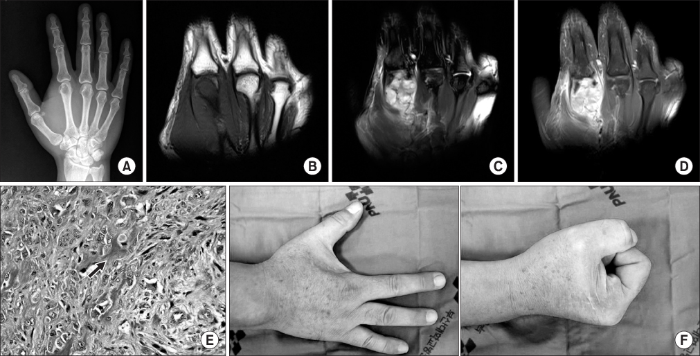
Figure 1 Osteosarcoma of the hand of a 56-year-old male patient. (A) Posteroanterior radiograph of the hand showed an ill-defined osteolytic lesion of the head of the second metacarpal bone, and no definite cortical destruction or soft tissue was found. (B) Coronal T1-weighted image showed a low signal intensity mass in the head of the second metacarpal bone and soft tissue. (C) Coronal T2-weighted image showed a high signal intensity mass in the head of the second metacarpal bone and soft tissue with cortical destruction, and the mass was expanding on the volar side. (D) Gadolinium-enhanced fat saturated T1-weighted image showed inhomogeneous contrast enhancement in the head of the second metacarpal bone and soft tissue. (E) Histologic feature showed a fibroblastic osteosarcoma, showing atypical stromal cells with malignant osteoid (black arrow) (H&E, ×400). (F) Gross appearance of the right hand four years after ray amputation of second finger showed excellent function and appearance.
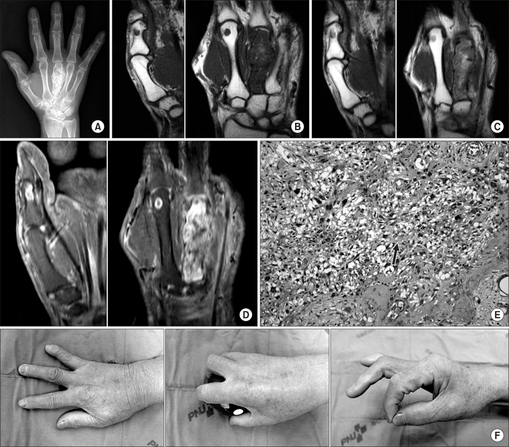
Figure 2 Osteosarcoma of the hand of a 73-year-old female patient. (A) Posteroanterior radiograph of the hand showed an endosteal expansile calcified mass in the entire third metacarpal bone suggesting as malignant transformation of enchondroma. (B-D) Magnetic resonance image showed 5 mm multifocal ring-like masses in the first proximal phalanx and head of the second metacarpal bone, and a large endosteal expansile mass with cortical destruction in the entire third metacarpal bone, suggesting malignant transformation of enchondroma. This mass showed low signal intensity in a T1-weighted image, intermediate signal intensity in a T2-weighted image, and inhomogeneous contrast enhancement in a gadolinium-enhanced fat saturated T1-weighted image. (E) Histologic feature showed an osteoblastic osteosarcoma, showing atypical stromal cells with malignant osteoid (black arrow) (H&E, ×200). (F) Gross appearance and radiographs of the right hand five years after ray amputation of the third finger showed excellent function and appearance.
Reference
-
1. Anninga JK, Picci P, Fiocco M, et al. Osteosarcoma of the hands and feet: a distinct clinico-pathological subgroup. Virchows Arch. 2013; 462:109–120.
Article2. Fowble VA, Pae R, Vitale A, Bryk E, Vigorita VJ. Case reports: osteosarcoma of the hand: one case and a literature review. Clin Orthop Relat Res. 2005; 440:255–261.3. Sforzo CR, Scarborough MT, Wright TW. Bone-forming tumors of the upper extremity and Ewing's sarcoma. Hand Clin. 2004; 20:303–315.
Article4. Okada K, Wold LE, Beabout JW, Shives TC. Osteosarcoma of the hand. A clinicopathologic study of 12 cases. Cancer. 1993; 72:719–725.
Article5. Clifford RH, Kelly AP Jr. Primary malignant tumors of the hand. Plast Reconstr Surg (1946). 1955; 15:227–232.
Article6. Larsson SE, Lorentzon R, Wedrén H, Boquist L. Osteosarcoma. A multifactorial clinical and histopathological study with special regard to therapy and survival. Acta Orthop Scand. 1978; 49:571–581.7. Kerin R. Metastatic tumors of the hand. A review of the literature. J Bone Joint Surg Am. 1983; 65:1331–1335.
Article8. Rosen G, Marcove RC, Caparros B, Nirenberg A, Kosloff C, Huvos AG. Primary osteogenic sarcoma: the rationale for preoperative chemotherapy and delayed surgery. Cancer. 1979; 43:2163–2177.
Article9. Papagelopoulos PJ, Galanis EC, Vlastou C, et al. Current concepts in the evaluation and treatment of osteosarcoma. Orthopedics. 2000; 23:858–867.
Article10. Posner MA. Ray transposition for central digital loss. J Hand Surg Am. 1979; 4:242–257.
Article
