Bone Reconstruction: Structural Allograft and Autograft
- Affiliations
-
- 1Department of Orthopedic Surgery, College of Medicine, The Catholic University of Korea, Seoul, Korea. ygchung@catholic.ac.kr
- KMID: 2185087
- DOI: http://doi.org/10.4055/jkoa.2015.50.6.462
Abstract
- Structural allograft or recycled autograft bone transplantation has been performed for reconstruction of bone defects caused by bone tumor resection. Knowledge regarding advantages and disadvantages of bone reconstruction using an allograft or recycled autograft, other alternatives such as reconstruction with tumor prosthesis, the understanding of biologic characteristics and fate of transplanted bones, functional results, and complications of reconstruction are important. The surgeon should also be accustomed to the major technical points of allograft or recycled autograft transplantation. Proper indication, selection of an appropriate allograft or recycled autograft, rigid fixation, accurate surgical skills, preventive measures of infection and efficient rehabilitation are necessary in order to obtain long term survival of grafted bones and good functional outcome. Here, I will discuss the bone reconstruction methods using structural allograft or recycled autograft transplantation after bone tumor resection and their clinical results.
MeSH Terms
Figure
-

Figure 1 (A) An anteroposterior radiograph of the right humerus of a 12-year-old patient in whom an osteochondral allograft reconstruction was performed after wide near total resection of the humerus involved with osteosarcoma. (B) A lateral radiograph of a patient with osteosarcoma at the midshaft of the right femur treated with intercalary allograft reconstruction. Dual plate fixation was performed for stability of the construct. (C) An anteroposterior radiograph of the right distal femur of a patient with osteosarcoma in whom resected bone was reconstructed with an allograft-prosthesis composite.

Figure 2 Unicondylar osteochondral allograft. A 60-year-old male patient with metastatic carcinoma involving the medial condyle of right distal femur (A) was managed with a unicondylar osteoarticular allograft (B). After resection of the medial hemicondyle (C), a size- and shape-matched allograft hemicondyle was inserted and fixated with plate and screws (D). (E) Postoperative radiograph showed a well reconstructed articular surface.
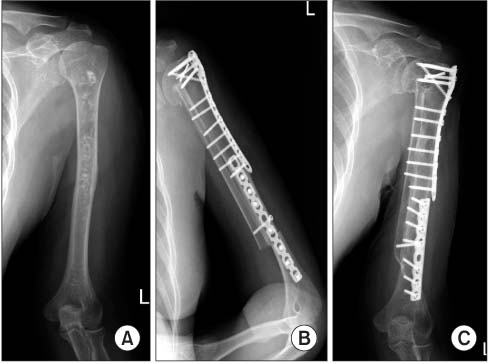
Figure 3 A 55-year-old male patient with chondrosarcoma at the left humerus (A) was managed with an intercalary allograft combined with an onlay type vascularized fibular graft (B). (C) Six year follow-up radiograph showed solid bone union at both proximal and distal host-allograft bone junctions (Courtesy of Dr. Joo-Yup Lee, Department of Orthopedic Surgery, The Catholic University of Korea, St. Vincent Hospital, Suwon; with permission).
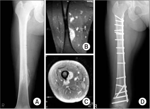
Figure 4 A 40-year-old female patient with parosteal osteosarcoma at the right distal femur (A-C) was managed with wide resection and reconstruction with a recycled pasteurized autograft combined with an inlay type vascularized fibular graft. (D) Twelve-year follow-up radiograph showed solid bone union at both proximal and distal host-allograft bone junctions. The patient recovered full limb function.
Reference
-
1. Lexer E. Substitution of whole or half joints from freshly amputated extremities by free plastic operation. Surg Gynecol Obstet. 1908; 6:601–607.2. Lexer E. Joint transplantation and arthroplasty. Surg Gynecol Obstet. 1925; 40:782–809.3. Curtiss PH Jr, Powell AE, Herndon CH. Immunological factors in homogenous-bone transplantation. II. The inability of homogenous rabbit bone to induce circulating antibodies in rabbits. J Bone Joint Surg Am. 1959; 41:1482–1488.4. Campbell CJ, Ishida H, Takahashi H, Kelly F. The transplantation of articular cartilage. An experimental study in dogs. J Bone Joint Surg Am. 1963; 45:1579–1592.5. Parrish FF. Allograft replacement of all or part of the end of a long bone following excision of a tumor. J Bone Joint Surg Am. 1973; 55:1–22.
Article6. Mankin HJ, Springfield DS, Gebhardt MC, Tomford WW. Current status of allografting for bone tumors. Orthopedics. 1992; 15:1147–1154.
Article7. Gebhardt MC, Roth YF, Mankin HJ. Osteoarticular allografts for reconstruction in the proximal part of the humerus after excision of a musculoskeletal tumor. J Bone Joint Surg Am. 1990; 72:334–345.
Article8. Mnaymneh W, Malinin T. Massive allografts in surgery of bone tumors. Orthop Clin North Am. 1989; 20:455–467.9. Muscolo DL, Petracchi LJ, Ayerza MA, Calabrese ME. Massive femoral allografts followed for 22 to 36 years Report of six cases. J Bone Joint Surg Br. 1992; 74:887–892.
Article10. Benedetti MG, Bonatti E, Malfitano C, Donati D. Comparison of allograft-prosthetic composite reconstruction and modular prosthetic replacement in proximal femur bone tumors: functional assessment by gait analysis in 20 patients. Acta Orthop. 2013; 84:218–223.11. Farid Y, Lin PP, Lewis VO, Yasko AW. Endoprosthetic and allograft-prosthetic composite reconstruction of the proximal femur for bone neoplasms. Clin Orthop Relat Res. 2006; 442:223–229.
Article12. Hazan EJ, Hornicek FJ, Tomford W, Gebhardt MC, Mankin HJ. The effect of adjuvant chemotherapy on osteoarticular allografts. Clin Orthop Relat Res. 2001; 385:176–181.
Article13. Muscolo DL, Ayerza MA, Aponte-Tinao LA, Abalo E, Farfalli G. Unicondylar osteoarticular allografts of the knee. J Bone Joint Surg Am. 2007; 89:2137–2142.
Article14. Bianchi G, Staals EL, Donati D, Mercuri M. The use of unicondylar osteoarticular allografts in reconstructions around the knee. Knee. 2009; 16:1–5.
Article15. Aponte-Tinao L, Farfalli GL, Ritacco LE, Ayerza MA, Muscolo DL. Intercalary femur allografts are an acceptable alternative after tumor resection. Clin Orthop Relat Res. 2012; 470:728–734.
Article16. Frisoni T, Cevolani L, Giorgini A, Dozza B, Donati DM. Factors affecting outcome of massive intercalary bone allografts in the treatment of tumours of the femur. J Bone Joint Surg Br. 2012; 94:836–841.
Article17. Farfalli GL, Aponte-Tinao L, Lopez-Millán L, Ayerza MA, Muscolo DL. Clinical and functional outcomes of tibial intercalary allografts after tumor resection. Orthopedics. 2012; 35:e391–e396.
Article18. Zehr RJ, Enneking WF, Scarborough MT. Allograft-prosthesis composite versus megaprosthesis in proximal femoral reconstruction. Clin Orthop Relat Res. 1996; 322:207–223.
Article19. Langlais F, Lambotte JC, Collin P, Thomazeau H. Long-term results of allograft composite total hip prostheses for tumors. Clin Orthop Relat Res. 2003; 414:197–211.
Article20. Farfalli GL, Aponte-Tinao LA, Ayerza MA, et al. Comparison between Constrained and Semiconstrained Knee Allograft-Prosthesis Composite Reconstructions. Sarcoma. 2013; 2013:489652.
Article21. Ogilvie CM, Crawford EA, Hosalkar HS, King JJ, Lackman RD. Long-term results for limb salvage with osteoarticular allograft reconstruction. Clin Orthop Relat Res. 2009; 467:2685–2690.
Article22. Muscolo DL, Ayerza MA, Aponte-Tinao LA, Ranalletta M. Use of distal femoral osteoarticular allografts in limb salvage surgery. J Bone Joint Surg Am. 2005; 87:2449–2455.
Article23. Muscolo DL, Ayerza MA, Farfalli G, Aponte-Tinao LA. Proximal tibia osteoarticular allografts in tumor limb salvage surgery. Clin Orthop Relat Res. 2010; 468:1396–1404.
Article24. DeGroot H, Donati D, Di Liddo M, Gozzi E, Mercuri M. The use of cement in osteoarticular allografts for proximal humeral bone tumors. Clin Orthop Relat Res. 2004; 427:190–197.
Article25. Kocher MS, Gebhardt MC, Mankin HJ. Reconstruction of the distal aspect of the radius with use of an osteoarticular allograft after excision of a skeletal tumor. J Bone Joint Surg Am. 1998; 80:407–419.
Article26. Bianchi G, Donati D, Staals EL, Mercuri M. Osteoarticular allograft reconstruction of the distal radius after bone tumour resection. J Hand Surg Br. 2005; 30:369–373.
Article27. Mankin HJ, Gebhardt MC, Jennings LC, Springfield DS, Tomford WW. Long-term results of allograft replacement in the management of bone tumors. Clin Orthop Relat Res. 1996; 324:86–97.
Article28. Donati D, Giacomini S, Gozzi E, et al. Allograft arthrodesis treatment of bone tumors: a two-center study. Clin Orthop Relat Res. 2002; 400:217–224.
Article29. Deijkers RL, Bloem RM, Hogendoorn PC, Verlaan JJ, Kroon HM, Taminiau AH. Hemicortical allograft reconstruction after resection of low-grade malignant bone tumours. J Bone Joint Surg Br. 2002; 84:1009–1014.
Article30. Manfrini M, Vanel D, De Paolis M, et al. Imaging of vascularized fibula autograft placed inside a massive allograft in reconstruction of lower limb bone tumors. AJR Am J Roentgenol. 2004; 182:963–970.
Article31. Capanna R, Bufalini C, Campanacci C. A new technique for reconstruction of large metadiaphyseal bone defects: a combined graft (allograft shell plus vascularized fibula). Orthop Traumatol. 1993; 2:159–177.32. Ozaki T, Hillmann A, Wuisman P, Winkelmann W. Reconstruction of tibia by ipsilateral vascularized fibula and allograft. 12 cases with malignant bone tumors. Acta Orthop Scand. 1997; 68:298–301.
Article33. Moran SL, Shin AY, Bishop AT. The use of massive bone allograft with intramedullary free fibular flap for limb salvage in a pediatric and adolescent population. Plast Reconstr Surg. 2006; 118:413–419.
Article34. Enneking WF, Campanacci DA. Retrieved human allografts: a clinicopathological study. J Bone Joint Surg Am. 2001; 83-A:971–986.35. Gebhardt MC, Flugstad DI, Springfield DS, Mankin HJ. The use of bone allografts for limb salvage in high-grade extremity osteosarcoma. Clin Orthop Relat Res. 1991; 270:181–196.
Article36. Choong PF. The role of allografts in tumour surgery. Acta Orthop Scand Suppl. 1997; 273:89–94.
Article37. Donati D, Di Liddo M, Zavatta M, et al. Massive bone allograft reconstruction in high-grade osteosarcoma. Clin Orthop Relat Res. 2000; 377:186–194.
Article38. Mankin HJ, Doppelt S, Tomford W. Clinical experience with allograft implantation The first ten years. Clin Orthop Relat Res. 1983; 174:69–86.39. Enneking WF, Dunham W, Gebhardt MC, Malawar M, Pritchard DJ. A system for the functional evaluation of reconstructive procedures after surgical treatment of tumors of the musculoskeletal system. Clin Orthop Relat Res. 1993; 286:241–246.
Article40. Innocenti M, Abed YY, Beltrami G, Delcroix L, Manfrini M, Capanna R. Biological reconstruction after resection of bone tumors of the proximal tibia using allograft shell and intramedullary free vascularized fibular graft: long-term results. Microsurgery. 2009; 29:361–372.
Article41. Mankin HJ, Hornicek FJ, Raskin KA. Infection in massive bone allografts. Clin Orthop Relat Res. 2005; 432:210–216.
Article42. Berrey BH Jr, Lord CF, Gebhardt MC, Mankin HJ. Fractures of allografts. Frequency, treatment, and end-results. J Bone Joint Surg Am. 1990; 72:825–833.
Article43. Lietman SA, Tomford WW, Gebhardt MC, Springfield DS, Mankin HJ. Complications of irradiated allograft in orthopaedic surgery. Clin Orthop Relat Res. 2000; 375:214–217.44. Sorger JI, Hornicek FJ, Zavatta M, et al. Allograft fractures revisited. Clin Orthop Relat Res. 2001; 382:66–74.
Article45. Delloye C, de Nayer P, Allington N, Munting E, Coutelier L, Vincent A. Massive bone allografts in large skeletal defects after tumor surgery: a clinical and microradiographic evaluation. Arch Orthop Trauma Surg. 1988; 107:31–41.
Article46. Zehr RJ, Enneking WF, Heare T, Liang TS. Fractures in large structural allografts. In : Complications of limb salvage: prevention, management and outcome: 6th International Symposium; 1991 Sep 8-11; Montreal, Canada.47. Vander Griend RA. The effect of internal fixation on the healing of large allografts. J Bone Joint Surg Am. 1994; 76:657–663.
Article48. Hornicek FJ, Gebhardt MC, Tomford WW, et al. Factors affecting nonunion of the allograft-host junction. Clin Orthop Relat Res. 2001; 382:87–98.
Article49. Kumta SM, Leung PC, Griffith JF, Roebuck DJ, Chow LT, Li CK. A technique for enhancing union of allograft to host bone. J Bone Joint Surg Br. 1998; 80:994–998.
Article50. Song WS, Kong CB, Jeon DG, Cho WH, Kim JR, Lee SY. Overlapping allograft in reconstructive surgery for malignant bone tumours in paediatric patients: a preliminary report. J Bone Joint Surg Br. 2011; 93:537–541.51. Dick HM, Strauch RJ. Infection of massive bone allografts. Clin Orthop Relat Res. 1994; 306:46–53.52. Lord CF, Gebhardt MC, Tomford WW, Mankin HJ. Infection in bone allografts. Incidence, nature, and treatment. J Bone Joint Surg Am. 1988; 70:369–376.
Article53. Henderson ER, Groundland JS, Pala E, et al. Failure mode classification for tumor endoprostheses: retrospective review of five institutions and a literature review. J Bone Joint Surg Am. 2011; 93:418–429.
Article54. Tomford WW, Starkweather RJ, Goldman MH. A study of the clinical incidence of infection in the use of banked allograft bone. J Bone Joint Surg Am. 1981; 63:244–248.
Article55. Ritacco LE, Seiler C, Farfalli GL, et al. Validity of an automatic measure protocol in distal femur for allograft selection from a three-dimensional virtual bone bank system. Cell Tissue Bank. 2013; 14:213–220.
Article56. Duffy GP, Wood MB, Rock MG, Sim FH. Vascularized free fibular transfer combined with autografting for the management of fracture nonunions associated with radiation therapy. J Bone Joint Surg Am. 2000; 82:544–554.
Article57. Chen WM, Chen TH, Huang CK, Chiang CC, Lo WH. Treatment of malignant bone tumours by extracorporeally irradiated autograft-prosthetic composite arthroplasty. J Bone Joint Surg Br. 2002; 84:1156–1161.
Article58. Jeon DG, Kim MS, Cho WH, Song WS, Lee SY. Pasteurized autograft-prosthesis composite for distal femoral osteosarcoma. J Orthop Sci. 2007; 12:542–549.
Article59. Kim HS, Kim KJ, Han I, Oh JH, Lee SH. The use of pasteurized autologous grafts for periacetabular reconstruction. Clin Orthop Relat Res. 2007; 464:217–223.
Article60. Manabe J, Ahmed AR, Kawaguchi N, Matsumoto S, Kuroda H. Pasteurized autologous bone graft in surgery for bone and soft tissue sarcoma. Clin Orthop Relat Res. 2004; 419:258–266.
Article61. Tsuchiya H, Nishida H, Srisawat P, et al. Pedicle frozen autograft reconstruction in malignant bone tumors. J Orthop Sci. 2010; 15:340–349.
Article62. Tsuchiya H, Wan SL, Sakayama K, Yamamoto N, Nishida H, Tomita K. Reconstruction using an autograft containing tumour treated by liquid nitrogen. J Bone Joint Surg Br. 2005; 87:218–225.
Article63. Yoshida T, Sakamoto A, Tsukamoto N, Nakayama K, Iwamoto Y. Establishment of an animal model of a pasteurized bone graft, with a preliminary analysis of muscle coverage or FGF-2 administration to the graft. J Orthop Surg Res. 2009; 4:31.
Article64. Sugiura H, Nishida Y, Nakashima H, Yamada Y, Tsukushi S, Yamada K. Evaluation of long-term outcomes of pasteurized autografts in limb salvage surgeries for bone and soft tissue sarcomas. Arch Orthop Trauma Surg. 2012; 132:1685–1695.
Article65. Spira E, Brenner HT, Lubin E. Extracorporeal irradiation of malignant bone tumors. In : Chapchal G, editor. Operative treatment of bone tumors. Stuttgart: Thieme;1970. p. 136–140.66. Hamaker RC. Irradiation autogenous mandibular grafts in primary reconstructions. Laryngoscope. 1981; 91:1031–1051.67. Kotb SZ, Mostafa MF. Recycling of extracorporeally irradiated autograft for malignant bone tumors: long-term follow-up. Ann Plast Surg. 2013; 71:493–499.
- Full Text Links
-
- Actions
-
Cited
- CITED
-
- Close
- Share
-
- Similar articles
-
- A Comparative Study of Autograft and Tutoplast Processed Allograft in Posterolateral Lumbar Spine Fusion
- Comparison of Clinical Results in Anterior Cruciate Ligament Reconstruction Using Bone-Patellar Tendon-Bone Autograft and Using Achilles Tendon Allograft
- Comparison Between Allograft Mixed with Local Bone and Autograft in Posterolateral Lumbar Fusion
- Graft Considerations for Successful Anterior Cruciate Ligament Reconstruction
- Clinical results of Anterior Cruciate Ligament(ACL) Reconstruction: Bone-patellar Tendon-bone Autograft Versus Allograft
