Congenital Absence of the Pericardium
- Affiliations
-
- 1Cardiovascular Center, Seoul National University Bundang Hospital, Seongnam, Korea. flammeus1@gmail.com
- 2Department of Internal Medicine, Seoul National University College of Medicine, Seoul, Korea.
- 3Department of Radiology, Seoul National University Bundang Hospital, Seongnam, Korea.
- KMID: 2177455
- DOI: http://doi.org/10.4250/jcu.2014.22.1.36
Abstract
- Congenital absence of the pericardium is a rare cardiac malformation and is most often asymptomatic. It is usually discovered as an incidental finding. Physical examination, chest radiography, and electrocardiogram are often unremarkable. Echocardiography provides valuable information, and sometimes computed tomography or magnetic resonance imaging is needed for subsequent confirmation.
MeSH Terms
Figure
-

Fig. 1 A: 12-lead electrocardiogram demonstrating normal sinus rhythm and right deviation of the heart axis. B: Chest radiograph demonstrating leftward position of the heart and the bulging contour of the left heart border. C: Echocardiography apical four chamber view: echocardiography showed a globe-shaped heart and bulbous ventricle. D: Chest CT revealed outpouching of the RV, which contacted the left chest wall (white arrows). E: Chest CT showed interposition of the lung between the aortic arch and pulmonary trunk (yellow arrow). LA: left atrium, LV: left ventricle, RA: right atrium, RV: right ventricle, Ao: aorta.

Fig. 2 A: 12-lead electrocardiogram demonstrating sinus rhythm with an incomplete right bundle branch block. B: Chest radiograph demonstrating leftward position of the heart, flattening of the left heart border, and a lucent area between the aorta and pulmonary artery (white arrow). C: Echocardiography apical four chamber view: left ventricular apex showed a swinging motion in diastole and systole. D: Chest CT revealed nonvisualization of the pericardium (yellow arrows) and left-ward displacement of the entire heart with mild right ventricular dilatation.
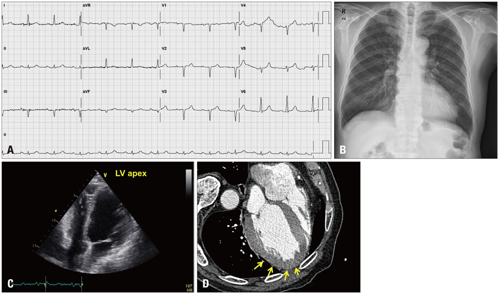
Fig. 3 A: Electrocardiogram demonstrating normal sinus rhythm with left deviation of the heart axis. B: Bulging contour of the left superior cardiac border. C: Apical four chamber view: echocardiography showed a laterally displaced left ventricular apex. D: The cardiac CT revealed absence of the pericardium at the left side of the heart (yellow arrows).
Reference
-
1. Abbas AE, Appleton CP, Liu PT, Sweeney JP. Congenital absence of the pericardium: case presentation and review of literature. Int J Cardiol. 2005; 98:21–25.
Article2. Centola M, Longo M, De Marco F, Cremonesi G, Marconi M, Danzi GB. Does echocardiography play a role in the clinical diagnosis of congenital absence of pericardium? A case presentation and a systematic review. J Cardiovasc Med (Hagerstown). 2009; 10:687–692.
Article3. D'Altorio R, Cano JY. Congenital abscence of the left pericardium detected by imaging of the lung: case report. J Nucl Med. 1977; 18:267–268.4. Eyileten Z, Arikbuka M, Yazicioğlu L, Ozyurda U. Left pericardial agenesis in a patient with sinus venosus type atrial septal defect. Anadolu Kardiyol Derg. 2007; 7:205–206.5. Lu C, Ridker PM. Echocardiographic diagnosis of congenital absence of the pericardium in a patient with VATER association defects. Clin Cardiol. 1994; 17:503–504.
Article6. Zakowski MF, Wright Y, Ricci A Jr. Pericardial agenesis and focal aplasia cutis in tetrasomy 12p (Pallister-Killian syndrome). Am J Med Genet. 1992; 42:323–325.
Article7. Boscherini B, Galasso C, Bitti ML. Abnormal face, congenital absence of the left pericardium, mental retardation, and growth hormone deficiency. Am J Med Genet. 1994; 49:111–113.
Article8. Sivrikoz MC, Durceylan E, Boztepe H, Birdane A. Congenital total absence of pericardium in a patient with left lung lower lobe bronchiectasis. Anadolu Kardiyol Derg. 2011; 11:81–83.
Article9. Lau KW, Ding ZP. Images in cardiovascular medicine. Partial pericardial defect. Circulation. 1998; 97:1992.10. Faridah Y, Julsrud PR. Congenital absence of pericardium revisited. Int J Cardiovasc Imaging. 2002; 18:67–73.11. Hoey ET, Yap KS, Darby MJ, Mankad K, Puppala S, Sivananthan MU. Complete left pericardial defect: evaluation with supine and decubitus dual source CT. J Cardiovasc Comput Tomogr. 2009; 3:417–419.
Article12. Hotouras A, Shahin Y. Pericardial agenesis: a rare cause of chest pain. Postgrad Med J. 2010; 86:380–381.
Article13. Gatzoulis MA, Munk MD, Merchant N, Van Arsdell GS, McCrindle BW, Webb GD. Isolated congenital absence of the pericardium: clinical presentation, diagnosis, and management. Ann Thorac Surg. 2000; 69:1209–1215.
Article
- Full Text Links
-
- Actions
-
Cited
- CITED
-
- Close
- Share
-
- Similar articles
-
- Congenital Complete Left-Sided Absence of the Pericardium Incidentally Identified in the Autopsy: A Case Report
- Incidentally Detected Pericardial Defect in a Patient with Pneumothorax as Confirmed on Video-Assisted Thoracoscopic Surgery
- Indentation in the Right Ventricle by an Incomplete Pericardium on 3-Dimensional Reconstructed Computed Tomography
- A Case of Congenital Pericardial Defect
- Congenital Left Atrial Appendage Aneurysm: A Case Report
