J Cardiovasc Ultrasound.
2012 Dec;20(4):218-219. 10.4250/jcu.2012.20.4.218.
A Case of Esophageal Achalasia Compressing Left Atrium Diagnosed by Echocardiography in Patient with Acute Chest Pain
- Affiliations
-
- 1Division of Cardiology, Severance Cardiovascular Hospital, Yonsei University College of Medicine, Seoul, Korea. grhong@yuhs.ac
- KMID: 2177414
- DOI: http://doi.org/10.4250/jcu.2012.20.4.218
Abstract
- No abstract available.
Keyword
Figure
-
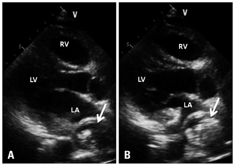
Fig. 1 A: Parasternal long axis view of transthoracic echocardiography showed round shape mass lesion compressing the left atrium (white arrow). B: After drinking a liquid containing carbon dioxide, the esophagus was filled with air contrast (white arrow). LA: left atrium, LV: left ventricle, RV: right ventricle.
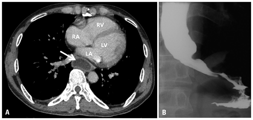
Fig. 2 A: Dilatation of the esophagus compressing the left atrium was revealed in chest computed tomography (white arrow). B: Esophagography with gastrografin identified achalasia. LA: left atrium, LV: left ventricle, RV: right ventricle, RA: right atrium.
Reference
-
1. D'Cruz IA, Feghali N, Gross CM. Echocardiographic manifestations of mediastinal masses compressing or encroaching on the heart. Echocardiography. 1994. 11:523–533.2. van Rooijen JM, van den Merkhof LF. Left atrial impression: a sign of extra-cardiac pathology. Eur J Echocardiogr. 2008. 9:661–664.
Article3. Goel K, Rishikant N, Chadha DS. Left atrial compression caused by achalasia. J Am Coll Cardiol. 2009. 54:955.
Article
- Full Text Links
-
- Actions
-
Cited
- CITED
-
- Close
- Share
-
- Similar articles
-
- A Case of Esophageal Carcinoma Following Esophagomyotomy for Achalasia
- A Case of Type II Achalasia Occurring in a Nonagenarian Diagnosed with Acute Food Impaction
- Diagnosis of Achalasia
- A Case of Esophageal Carcinoma in a Patient with Primary Achalasia
- A Case of Vigorous Achalasia Treated by Combined Treatments with Pneumatic Balloon Dilatation and Botulinum Toxin Injection
