Isolated Left Ventricular Noncompaction with a Congenital Aneurysm Presenting with Recurrent Embolism
- Affiliations
-
- 1Department of Internal Medicine, Gyeongsang National University Hospital, Jinju, Korea. parkjrang@gmail.com
- 2Department of Radiology, Gyeongsang National University Hospital, Jinju, Korea.
- KMID: 2177375
- DOI: http://doi.org/10.4250/jcu.2012.20.2.103
Abstract
- Isolated left ventricular noncompaction (LVNC) is a rare disorder caused by embryonic arrest of compaction. LVNC is sometimes associated with other congenital cardiac disorders; however, there have been few reports of its coexistence with a left ventricular aneurysm. A 40-year-old woman was admitted to our hospital for renal infarction. She had a history of embolic cerebral infarction 10 years ago. Transthoracic echocardiography showed prominent trabeculae and deep intertrabecular recesses which are filled with blood from the left ventricular (LV) cavity. A thrombus in the akinetic apical wall was confirmed by contrast echocardiography. Using cardiac computed tomography and magnetic resonance imaging, we rejected a possible diagnosis of suspicion of coronary artery disease. She was diagnosed LVNC with a thrombus in apical aneurysm. Here, we report the first patient in Korea known to have LVNC accompanying LV congenital aneurysm presenting with recurrent embolism.
MeSH Terms
Figure
-

Fig. 1 Transthoracic echocardiography. (A) A modified 4-chamber view showing an apical aneurysm (An) and prominent trabeculations (arrows) on the basal to mid-lateral wall. (B) Color Doppler image showing blood flow in the recess between trabeculae. (C and D) Apical level of short axis view showing trabeculations and perfusion in intertrabecular recesses on color Doppler imaging.
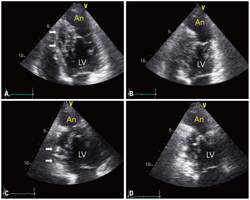
Fig. 2 Apical 4-chamber view (A and B) and apical 2-chamber view (C and D) in transthoracic echocardiography, showing increased trabeculations of the septal and inferior wall (arrows) and an apical aneurysm (An). The apical aneurysm of akinetic motion was defined at end-diastole (A and C) and end-systole (B and D). LV: left ventricle.
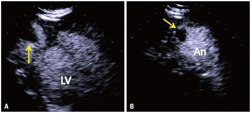
Fig. 3 Apical focusing image of apical long axis view (A) and apical short axis view (B) in contrast echocardiography, showing a thrombus (arrow) in the apical aneurysm. LV: left ventricle, An: aneurysm.

Fig. 4 Coronary computed tomography angiography, showing normal coronary artery (A), thrombus (arrow) in an apical aneurysm (B), and a thick noncompacted layer (C). LAD: left anterior descending artery, LCX: left circumflex artery, RCA: right coronary artery, LA: left atrium, LV: left ventricle, RV: right ventricle.
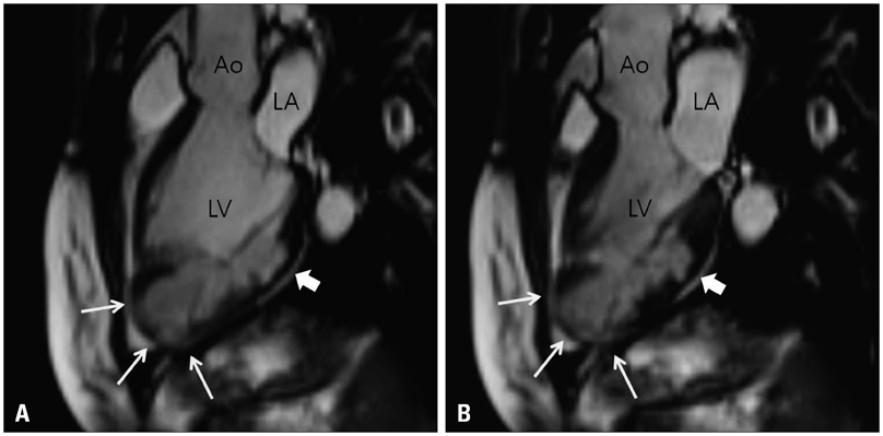
Fig. 5 Cardiac magnetic resonance imaging, showing a thin compacted layer preserving contractility (thick arrow) and an akinetic aneurysm (thin arrows) at end-diastole (A) and end-systole (B). Ao: aorta, LA: left atrium, LV: left ventricle.
Reference
-
1. Chin TK, Perloff JK, Williams RG, Jue K, Mohrmann R. Isolated noncompaction of left ventricular myocardium. A study of eight cases. Circulation. 1990. 82:507–513.
Article2. Ritter M, Oechslin E, Sütsch G, Attenhofer C, Schneider J, Jenni R. Isolated noncompaction of the myocardium in adults. Mayo Clin Proc. 1997. 72:26–31.
Article3. Burke A, Mont E, Kutys R, Virmani R. Left ventricular noncompaction: a pathological study of 14 cases. Hum Pathol. 2005. 36:403–411.
Article4. Ichida F. Left ventricular noncompaction. Circ J. 2009. 73:19–26.
Article5. Attenhofer Jost CH, Connolly HM, Warnes CA, O'leary P, Tajik AJ, Pellikka PA, Seward JB. Noncompacted myocardium in Ebstein's anomaly: initial description in three patients. J Am Soc Echocardiogr. 2004. 17:677–680.
Article6. Betrián Blasco P, Gallardo Agromayor E. Ebstein's anomaly and left ventricular noncompaction association. Int J Cardiol. 2007. 119:264–265.
Article7. Biagini E, Ragni L, Ferlito M, Pasquale F, Lofiego C, Leone O, Rocchi G, Perugini E, Zagnoni S, Branzi A, Picchio FM, Rapezzi C. Different types of cardiomyopathy associated with isolated ventricular noncompaction. Am J Cardiol. 2006. 98:821–824.
Article8. Oechslin EN, Attenhofer Jost CH, Rojas JR, Kaufmann PA, Jenni R. Long-term follow-up of 34 adults with isolated left ventricular noncompaction: a distinct cardiomyopathy with poor prognosis. J Am Coll Cardiol. 2000. 36:493–500.
Article9. Oechslin E, Jenni R. Left ventricular non-compaction revisited: a distinct phenotype with genetic heterogeneity? Eur Heart J. 2011. 32:1446–1456.
Article10. Sato Y, Matsumoto N, Yoda S, Inoue F, Kunimoto S, Fukamizu S, Tani S, Takayama T, Tokai K, Kasamaki Y, Saito S, Uchiyama T, Koyama Y. Left ventricular aneurysm associated with isolated noncompaction of the ventricular myocardium. Heart Vessels. 2006. 21:192–194.
Article11. Ionescu CN, Turcot D. Left ventricular noncompaction and aneurysm revealed by left ventriculography. Catheter Cardiovasc Interv. 2011. [Epub ahead of print].
Article12. Yun H, Zeng MS, Jin H, Yang S. Isolated noncompaction of ventricular myocardium: a magnetic resonance imaging study of 11 patients. Korean J Radiol. 2011. 12:686–692.
Article13. Maron BJ, Towbin JA, Thiene G, Antzelevitch C, Corrado D, Arnett D, Moss AJ, Seidman CE, Young JB. American Heart Association. Council on Clinical Cardiology, Heart Failure and Transplantation Committee. Quality of Care and Outcomes Research and Functional Genomics and Translational Biology Interdisciplinary Working Groups. Council on Epidemiology and Prevention. Contemporary definitions and classification of the cardiomyopathies: an American Heart Association Scientific Statement from the Council on Clinical Cardiology, Heart Failure and Transplantation Committee; Quality of Care and Outcomes Research and Functional Genomics and Translational Biology Interdisciplinary Working Groups; and Council on Epidemiology and Prevention. Circulation. 2006. 113:1807–1816.
Article14. Elliott P, Andersson B, Arbustini E, Bilinska Z, Cecchi F, Charron P, Dubourg O, Kühl U, Maisch B, McKenna WJ, Monserrat L, Pankuweit S, Rapezzi C, Seferovic P, Tavazzi L, Keren A. Classification of the cardiomyopathies: a position statement from the European Society Of Cardiology Working Group on Myocardial and Pericardial Diseases. Eur Heart J. 2008. 29:270–276.
Article15. Jenni R, Oechslin E, Schneider J, Attenhofer Jost C, Kaufmann PA. Echocardiographic and pathoanatomical characteristics of isolated left ventricular non-compaction: a step towards classification as a distinct cardiomyopathy. Heart. 2001. 86:666–671.
Article
- Full Text Links
-
- Actions
-
Cited
- CITED
-
- Close
- Share
-
- Similar articles
-
- Noncompaction of Ventricular Myocardium Involving the Right Ventricle
- A case of isolated noncompaction of the ventricular myocardium in an elderly patient
- Stroke in a Young Individual with Left Ventricular Noncompaction and Left Atrium Standstill
- A Case of Congenital Left Ventricular Aneurysm in an Elderly Woman
- Isolated Right Ventricular Noncompaction Accompanied by Right Ventricular Failure
