An assessment of template-guided implant surgery in terms of accuracy and related factors
- Affiliations
-
- 1Department of Oral and Maxillofacial Surgery, Seoul Asan Medical Center, Seoul, Republic of Korea.
- 2Department of Prosthodontics, Ewha Womans University, School of Medicine, Seoul, Republic of Korea.
- 3Department of Oral and Maxillofacial Surgery, Seoul National University Dental Hospital, Seoul, Republic of Korea. omsljh@gmail.com
- 4Department of Prosthodontics, Seoul National University Dental Hospital, Seoul, Republic of Korea.
- KMID: 2176536
- DOI: http://doi.org/10.4047/jap.2013.5.4.440
Abstract
- PURPOSE
Template-guided implant therapy has developed hand-in-hand with computed tomography (CT) to improve the accuracy of implant surgery and future prosthodontic treatment. In our present study, the accuracy and causative factors for computer-assisted implant surgery were assessed to further validate the stable clinical application of this technique.
MATERIALS AND METHODS
A total of 102 implants in 48 patients were included in this study. Implant surgery was performed with a stereolithographic template. Pre- and post-operative CTs were used to compare the planned and placed implants. Accuracy and related factors were statistically analyzed with the Spearman correlation method and the linear mixed model. Differences were considered to be statistically significant at P< or =.05.
RESULTS
The mean errors of computer-assisted implant surgery were 1.09 mm at the coronal center, 1.56 mm at the apical center, and the axis deviation was 3.80degrees. The coronal and apical errors of the implants were found to be strongly correlated. The errors developed at the coronal center were magnified at the apical center by the fixture length. The case of anterior edentulous area and longer fixtures affected the accuracy of the implant template.
CONCLUSION
The control of errors at the coronal center and stabilization of the anterior part of the template are needed for safe implant surgery and future prosthodontic treatment.
Keyword
MeSH Terms
Figure
-
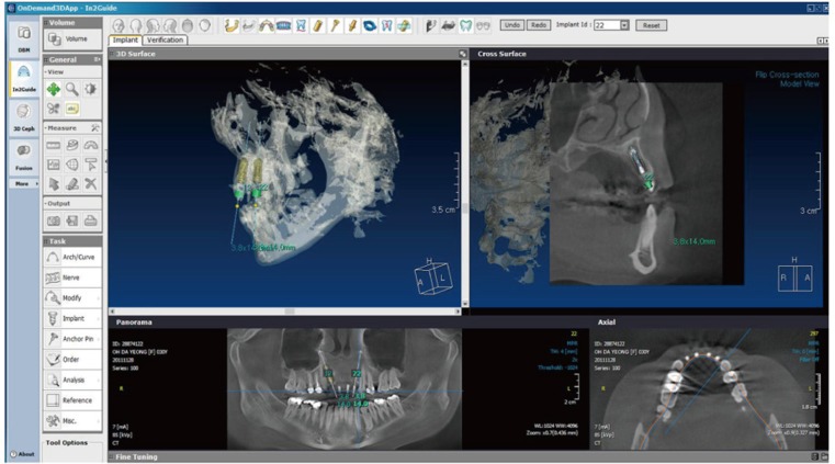
Fig. 1 The location and angulation of the implant were determined through the use of planning software and virtual surgery by considering the critical anatomical structures and the future dental prostheses.
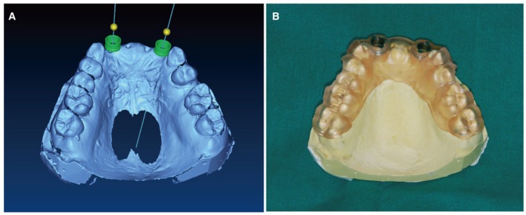
Fig. 2 The information from the planned fixture was used to create the stereolithographic implant template. (A) Completion of planning of implant surgery on 3D software, (B) Stereolithographic template for computer-assisted implant surgery.
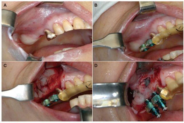
Fig. 3 Surgical procedure for template-guided implant surgery. (A) Preparation of operation field, (B) Setting of surgical template in the oral cavity, (C) Flap elevation for sequential drilling, (D) Implantation of fixture guided by the template.
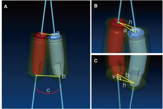
Fig. 4 (A) Comparison of the planned and placed implants by superimposition of the pre- and postoperative images, (B) Disassembly of the total errors into horizontal and vertical directions at the coronal center, (C) Disassembly of the total errors into horizontal and vertical directions at the apical center.a: coronal distance, b: apical distance, c: axis deviation, h: horizontal deviation, v: vertical deviation.
Cited by 2 articles
-
Accuracy of a direct drill-guiding system with minimal tolerance of surgical instruments used for implant surgery: a prospective clinical study
Du-Hyeong Lee, Seo-Young An, Min-Ho Hong, Kyoung-Bae Jeon, Kyu-Bok Lee
J Adv Prosthodont. 2016;8(3):207-213. doi: 10.4047/jap.2016.8.3.207.A 10-year follow-up study on clinical outcomes of dental implant rehabilitation using surgical guide
Haoyun Li, Mi Young Eo, Kezia Rachellea Mustakim, Soung Min Kim
J Korean Assoc Oral Maxillofac Surg. 2024;50(2):70-79. doi: 10.5125/jkaoms.2024.50.2.70.
Reference
-
1. Besimo C, Lambrecht JT, Nidecker A. Dental implant treatment planning with reformatted computed tomography. Dentomaxillofac Radiol. 1995; 24:264–267. PMID: 9161173.
Article2. Fortin T, Coudert JL, Champleboux G, Sautot P, Lavallée S. Computer-assisted dental implant surgery using computed tomography. J Image Guid Surg. 1995; 1:53–58. PMID: 9079427.
Article3. Jacobs R, Adriansens A, Naert I, Quirynen M, Hermans R, Van Steenberghe D. Predictability of reformatted computed tomography for pre-operative planning of endosseous implants. Dentomaxillofac Radiol. 1999; 28:37–41. PMID: 10202477.
Article4. Fortin T, Champleboux G, Bianchi S, Buatois H, Coudert JL. Precision of transfer of preoperative planning for oral implants based on cone-beam CT-scan images through a robotic drilling machine. Clin Oral Implants Res. 2002; 13:651–656. PMID: 12519341.
Article5. Ozan O, Turkyilmaz I, Ersoy AE, McGlumphy EA, Rosenstiel SF. Clinical accuracy of 3 different types of computed tomography-derived stereolithographic surgical guides in implant placement. J Oral Maxillofac Surg. 2009; 67:394–401. PMID: 19138616.
Article6. Jacobs R, Adriansens A, Verstreken K, Suetens P, van Steenberghe D. Predictability of a three-dimensional planning system for oral implant surgery. Dentomaxillofac Radiol. 1999; 28:105–111. PMID: 10522199.
Article7. Arisan V, Karabuda ZC, Ozdemir T. Accuracy of two stereolithographic guide systems for computer-aided implant placement: a computed tomography-based clinical comparative study. J Periodontol. 2010; 81:43–51. PMID: 20059416.8. Cassetta M, Di Mambro A, Giansanti M, Stefanelli LV, Cavallini C. The intrinsic error of a stereolithographic surgical template in implant guided surgery. Int J Oral Maxillofac Surg. 2013; 42:264–275. PMID: 22789635.
Article9. D'haese J, Van De Velde T, Elaut L, De Bruyn H. A prospective study on the accuracy of mucosally supported stereolithographic surgical guides in fully edentulous maxillae. Clin Implant Dent Relat Res. 2012; 14:293–303. PMID: 19906267.10. Park C, Raigrodski AJ, Rosen J, Spiekerman C, London RM. Accuracy of implant placement using precision surgical guides with varying occlusogingival heights: an in vitro study. J Prosthet Dent. 2009; 101:372–381. PMID: 19463664.
Article11. Stumpel LJ. Deformation of stereolithographically produced surgical guides: an observational case series report. Clin Implant Dent Relat Res. 2012; 14:442–453. PMID: 20156227.
Article12. Valente F, Schiroli G, Sbrenna A. Accuracy of computer-aided oral implant surgery: a clinical and radiographic study. Int J Oral Maxillofac Implants. 2009; 24:234–242. PMID: 19492638.13. Vercruyssen M, Jacobs R, Van Assche N, van Steenberghe D. The use of CT scan based planning for oral rehabilitation by means of implants and its transfer to the surgical field: a critical review on accuracy. J Oral Rehabil. 2008; 35:454–474. PMID: 18429973.
Article14. Di Giacomo GA, Cury PR, de Araujo NS, Sendyk WR, Sendyk CL. Clinical application of stereolithographic surgical guides for implant placement: preliminary results. J Periodontol. 2005; 76:503–507. PMID: 15857088.15. Vrielinck L, Politis C, Schepers S, Pauwels M, Naert I. Image-based planning and clinical validation of zygoma and pterygoid implant placement in patients with severe bone atrophy using customized drill guides. Preliminary results from a prospective clinical follow-up study. Int J Oral Maxillofac Surg. 2003; 32:7–14. PMID: 12653226.
Article16. Lee JH, Kim MJ, Kim SM, Kwon OH, Kim YK. The 3D CT superimposition method using image fusion based on the maximum mutual information algorithm for the assessment of oral and maxillofacial surgery treatment results. Oral Surg Oral Med Oral Pathol Oral Radiol. 2012; 114:167–174. PMID: 22776729.
Article17. Verstreken K, Van Cleynenbreugel J, Marchal G, Naert I, Suetens P, van Steenberghe D. Computer-assisted planning of oral implant surgery: a three-dimensional approach. Int J Oral Maxillofac Implants. 1996; 11:806–810. PMID: 8990645.18. Fortin T, Camby E, Alik M, Isidori M, Bouchet H. Panoramic images versus three-dimensional planning software for oral implant planning in atrophied posterior maxillary: a clinical radiological study. Clin Implant Dent Relat Res. 2013; 15:198–204. PMID: 21477064.
Article19. Cassetta M, Stefanelli LV, Giansanti M, Calasso S. Accuracy of implant placement with a stereolithographic surgical template. Int J Oral Maxillofac Implants. 2012; 27:655–663. PMID: 22616060.20. Sarment DP, Sukovic P, Clinthorne N. Accuracy of implant placement with a stereolithographic surgical guide. Int J Oral Maxillofac Implants. 2003; 18:571–577. PMID: 12939011.21. Kramer FJ, Baethge C, Swennen G, Rosahl S. Navigated vs. conventional implant insertion for maxillary single tooth replacement. Clin Oral Implants Res. 2005; 16:60–68. PMID: 15642032.
Article22. Choi M, Romberg E, Driscoll CF. Effects of varied dimensions of surgical guides on implant angulations. J Prosthet Dent. 2004; 92:463–469. PMID: 15523335.
Article
- Full Text Links
-
- Actions
-
Cited
- CITED
-
- Close
- Share
-
- Similar articles
-
- A survey of the satisfaction of patients who have undergone implant surgery with and without employing a computer-guided implant surgical template
- An assessment of accuracy of half-guided implant surgery using implant surgical guide: A case report
- Accuracy of the CT guided implant template by using an intraoral scanner according to the edentulous distance
- A Survey of Surgeons' Satisfactory Level on Using Zero-Setup Implant Surgical Guide System
- A Survey of Surgeons' Satisfactory Level on Using Zero-Setup Implant Surgical Guide System
