Stress distribution of oval and circular fiber posts in amandibular premolar: a three-dimensional finite element analysis
- Affiliations
-
- 1Department of Endodontics, Faculty of Dentistry, Erciyes University, Kayseri, Turkey. ozgurer@erciyes.edu.tr
- 2Department Prosthodontics, Faculty of Dentistry, Erciyes University, Kayseri, Turkey.
- 3Department of Mechanical Engineering, Faculty of Engineering, Erciyes University, Kayseri, Turkey.
- KMID: 2176535
- DOI: http://doi.org/10.4047/jap.2013.5.4.434
Abstract
- PURPOSE
The aim of the present study was to evaluate the effects of posts with different morphologies on stress distribution in an endodontically treated mandibular premolar by using finite element models (FEMs).
MATERIALS AND METHODS
A mandibular premolar was modeled using the ANSYS software program. Two models were created to represent circular and oval fiber posts in this tooth model. An oblique force of 300 N was applied at an angle of 45degrees to the occlusal plane and oriented toward the buccal side. von Mises stress was measured in three regions each for oval and circular fiber posts.
RESULTS
FEM analysis showed that the von Mises stress of the circular fiber post (426.81 MPa) was greater than that of the oval fiber post (346.34 MPa). The maximum distribution of von Mises stress was in the luting agent in both groups. Additionally, von Mises stresses accumulated in the coronal third of root dentin, close to the post space in both groups.
CONCLUSION
Oval fiber posts are preferable to circular fiber posts in oval-shaped canals given the stress distribution at the post-dentin interface.
MeSH Terms
Figure
-
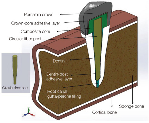
Fig. 1 Geometry of the model for circular fiber post.
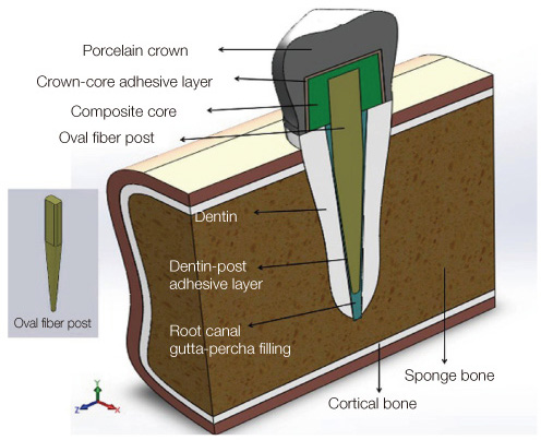
Fig. 2 Geometry of the model for oval fiber post.
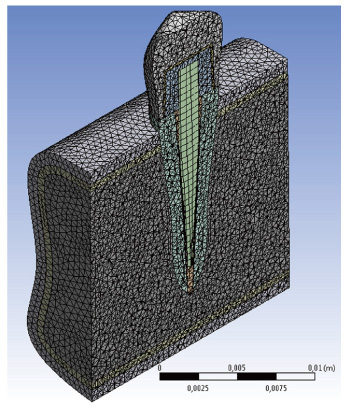
Fig. 3 The meshed model had 262,160 elements and 401,161 nodes for oval fiber post and 263,443 elements and 402,608 nodes for circular fiber post.
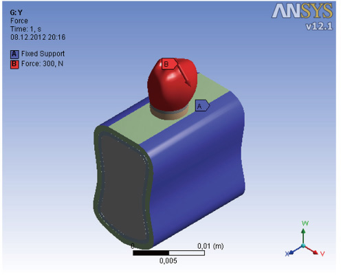
Fig. 4 Model loading conditions. The boundary condition of the model was a fixed support at the surface of the gingiva.

Fig. 5 von Mises stresses measuring from three regions.
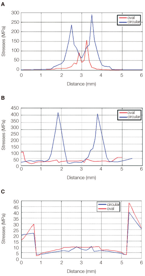
Fig. 6 (A) von Mises stresses measured along the apical extent of the post (post tip), (B) von Mises stresses measured combination of the core-root dentin interface from the labial to the palatal side, (C) von Mises stresses measured 3 mm above from the b point from the labial to the palatal side.
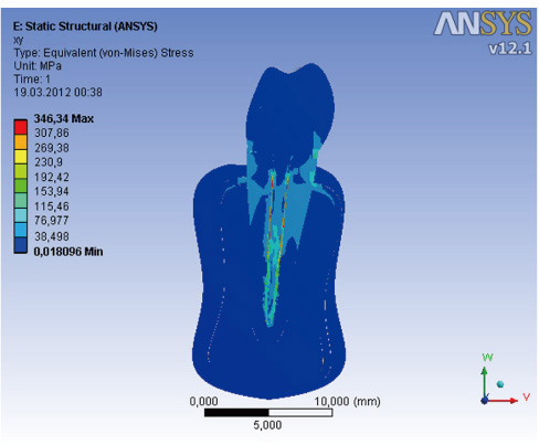
Fig. 7 von Mises stresses for oval fiber post.
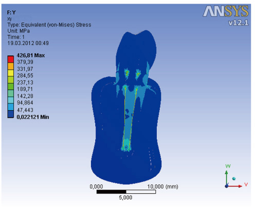
Fig. 8 von Mises stresses for circular fiber post.
Reference
-
1. Hess D, Solomon E, Spears R, He J. Retreatability of a bioceramic root canal sealing material. J Endod. 2011; 37:1547–1549.2. Cecchin D, de Almeida JF, Gomes BP, Zaia AA, Ferraz CC. Effect of chlorhexidine and ethanol on the durability of the adhesion of the fiber post relined with resin composite to the root canal. J Endod. 2011; 37:678–683.3. Faria-e-Silva AL, Pedrosa-Filho Cde F, Menezes Mde S, Silveira DM, Martins LR. Effect of relining on fiber post retention to root canal. J Appl Oral Sci. 2009; 17:600–604.4. Kremeier K, Fasen L, Klaiber B, Hofmann N. Influence of endodontic post type (glass fiber, quartz fiber or gold) and luting material on push-out bond strength to dentin in vitro. Dent Mater. 2008; 24:660–666.5. Giachetti L, Scaminaci Russo D, Baldini M, Bertini F, Steier L, Ferrari M. Push-out strength of translucent fibre posts cemented using a dual-curing technique or a light-curing self-adhering material. Int Endod J. 2012; 45:249–256.6. Muñoz C, Llena C, Forner L. Oval fiber posts do not improve adaptation to oval-shaped canal walls. J Endod. 2011; 37:1386–1389.7. Coniglio I, Magni E, Cantoro A, Goracci C, Ferrari M. Push-out bond strength of circular and oval-shaped fiber posts. Clin Oral Investig. 2011; 15:667–672.8. Coniglio I, Garcia-Godoy F, Magni E, Carvalho CA, Ferrari M. Resin cement thickness in oval-shaped canals: oval vs. circular fiber posts in combination with different tips/drills for post space preparation. Am J Dent. 2009; 22:290–294.9. Coniglio I, Carvalho CA, Magni E, Cantoro A, Ferrari M. Post space debridement in oval-shaped canals: the use of a new ultrasonic tip with oval section. J Endod. 2008; 34:752–755.10. Clark DJ. Shaping and restoring ovoid canal systems. Endod ther. 2005; 5:9–13.11. Boschian Pest L, Guidotti S, Pietrabissa R, Gagliani M. Stress distribution in a post-restored tooth using the three-dimensional finite element method. J Oral Rehabil. 2006; 33:690–697.12. Hsu ML, Chen CS, Chen BJ, Huang HH, Chang CL. Effects of post materials and length on the stress distribution of endodontically treated maxillary central incisors: a 3D finite element analysis. J Oral Rehabil. 2009; 36:821–830.13. Martínez-Insua A, da Silva L, Rilo B, Santana U. Comparison of the fracture resistances of pulpless teeth restored with a cast post and core or carbon-fiber post with a composite core. J Prosthet Dent. 1998; 80:527–532.14. Maceri F, Martignoni M, Vairo G. Mechanical behaviour of endodontic restorations with multiple prefabricated posts: a finite-element approach. J Biomech. 2007; 40:2386–2398.15. de Castro Albuquerque R, Polleto LT, Fontana RH, Cimini CA. Stress analysis of an upper central incisor restored with different posts. J Oral Rehabil. 2003; 30:936–943.16. Pierrisnard L, Bohin F, Renault P, Barquins M. Coronoradicular reconstruction of pulpless teeth: a mechanical study using finite element analysis. J Prosthet Dent. 2002; 88:442–448.17. Eskitaşcioğlu G, Belli S, Kalkan M. Evaluation of two post core systems using two different methods (fracture strength test and a finite elemental stress analysis). J Endod. 2002; 28:629–633.18. Pegoretti A, Fambri L, Zappini G, Bianchetti M. Finite element analysis of a glass fibre reinforced composite endodontic post. Biomaterials. 2002; 23:2667–2682.19. Aggarwal S, Garg V. Finite element analysis of stress concentration in three popular brands of fiber posts systems used for maxillary central incisor teeth. J Conserv Dent. 2011; 14:293–296.
- Full Text Links
-
- Actions
-
Cited
- CITED
-
- Close
- Share
-
- Similar articles
-
- Evaluation of the resin cement thicknesses and push-out bond strengths of circular and oval fiber posts in oval-shapes canals
- Finite element analysis of the influence of esthetic posts on incisors
- Effects of occlusal load on the cervical stress distribution: A three-dimensional finite element study
- A three-dimensional finite element analysis of obturator prosthesis for edentulous maxilla
- THREE DIMENSIONAL FINITE ELEMENT ANALYSIS ON THE MANDIBULAR CANTILEVERED PROSTHESIS SUPPORTED BY IMPLANTS
