Effects of occlusal load on the cervical stress distribution: A three-dimensional finite element study
- Affiliations
-
- 1Department of Conservative dentistry, College of Dentistry, Pusan National University, Korea. jeongkil@pusan.ac.kr
- 2Department of Mechanical design engineering, College of Engineering, Pusan National University, Korea.
- KMID: 1986885
- DOI: http://doi.org/10.5395/JKACD.2006.31.6.427
Abstract
- The objective of this study was to investigate the effects of various occlusal loads on the stress distribution of the buccal cervical region of a normal maxillary second premolar, using a three dimensional finite element analysis (3D FEA). After 3D FE modeling of maxillary second premolar, a static load of 500N of three load cases was applied. Stress analysis was performed using ANSYS (Swanson Analysis Systems, Inc., Houston, USA). The maximum principal stresses and minimum principal stresses were sampled at thirteen nodal points in the buccal cervical enamel for each four horizontal planes, 1.0 mm above CEJ, 0.5 mm above CEJ, CEJ, 0.5 mm under CEJ. The results were as follows 1. The peak stress was seen at the cervical enamel surface of the mesiobuccal line angle area, asymmetrically. 2. The values of compressive stresses were within the range of the failure stress of enamel. But the values of tensile stresses exceeded the range of the failure stress of enamel. 3. The tensile stresses from the perpendicular load at the buccal incline of palatal cusp may be shown to be the primary etiological factors of the NCCLs.
Keyword
Figure
-
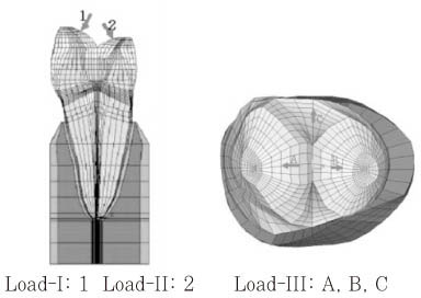
Figure 1 Three load conditions of 3D FE model.

Figure 2 Principal stress distribution of Load-I,II,III (Upper lines; tensile stress distributions Lower lines; compressive stress distributions).

Figure 3 Principal stresses of Level A,B,C,D of three load conditions.
Cited by 2 articles
-
Stress distribution of endodontically treated maxillary second premolars restored with different methods: Three-dimensional finite element analysis
Dong-Yeol Lim, Hyeon-Cheol Kim, Bock Hur, Kwang-Hoon Kim, Kwon Son, Jeong-Kil Park
J Korean Acad Conserv Dent. 2009;34(1):69-79. doi: 10.5395/JKACD.2009.34.1.069.Effect of restoration type on the stress distribution of endodontically treated maxillary premolars; Three-dimensional finite element study
Heun-Sook Jung, Hyeon-Cheol Kim, Bock Hur, Kwang-Hoon Kim, Kwon Son, Jeong-Kil Park
J Korean Acad Conserv Dent. 2009;34(1):8-19. doi: 10.5395/JKACD.2009.34.1.008.
Reference
-
1. Rees JS. A review of the biomechanics of abfraction. Eur J Prosthodont Restor Dent. 2000. 8(4):139–144.2. Lee WC, Eakle WS. Stress-induced cervical lesions: Review of advances on the past 10 years. J Prosthet Dent. 1996. 75:487–494.3. Grippo JO. Abfractions: A new classification of hard tissue lesions of teeth. J Esthet Dent. 1991. 3(1):14–19.
Article4. Rees JS, Hammadeh M. Undermining of enamel as a mechanism of abfraction lesion formation: A finite element study. Eur J Oral Sci. 2004. 112:347–352.
Article5. Lambrechts P, Braem M, Vanherle G. Evaluation of clinical performance for poster composite resins and dentin adhesives. Oper Dent. 1987. 12:53–78.6. Khan F, Young WG, Shahabi S, Daley TJ. Dental cervical lesions associated with occlusal erosion and attrition. Aust Dent J. 1999. 44:176–186.
Article7. Lee WC, Eakle WS. Possible role of the tensile stress in the etiology of cervical erosive lesions of teeth. J Prosthet Dent. 1984. 52(3):374–380.
Article8. Burke FJ, Whitehead SA, McCaughey AD. Contemporary concepts in the pathogenesis of the class V non-carious lesion. Dent update. 1995. 22(1):28–32.9. Aw TC, Lepe X, Johnson GH, Mancl L. Characteristics of noncarious cervical lesions. J Am Dent Assoc. 2002. 133:725–733.
Article10. Selna LG, Shillingdurg HT, Kerr PA. Finite element analysis of dental structures -axisymmetric and plane stress idealizations. J Biomed Mater Res. 1975. 9:237–252.
Article11. Yettram AL, Wright KW, Pickard HM. Finite element stress analysis of the crowns of normal and restored teeth. J Dent Res. 1976. 55(6):1004–1011.
Article12. Goel VK, Khera SC, Ralston JL, Chang KH. Stresses at the dentinoenamel junction of human teeth-A finite element investigation. J Prosthet Dent. 1991. 66:451–459.
Article13. Palamara D, Palamara JEA, Tyas MJ, Messer HH. Strain patterns in cervical enamel of teeth subjected to occlusal loading. Dent Mater. 2000. 16:412–419.
Article14. Rees JS, Hammadeh M, Jagger DC. Abfraction lesion formation in maxillary incisors, canines and premolars: A finite element study. Eur J Oral Sci. 2003. 111:149–154.
Article15. Tanaka M, Naito T, Yokota M, Kohno M. Finite element analysis of the possible mechanism of cervical lesion formation by occlusal force. J Oral Rehabil. 2003. 30:60–67.
Article16. Geramy A, Sharafoddin F. Abfraction: 3D analysis by means of the finite element method. Quintessence Int. 2003. 34:526–533.17. Katona TR, Winkler MM. Stress analysis of a bulk-filled Class V light-cured composite restoration. J Dent Res. 1994. 73(8):1470–1477.
Article18. Lindehe J, Karring T. Schluger S, Yuodelis R, Page RC, Johnson RH, editors. The anatomy of the periodontium. Textbook of Clinical Periodontology. 1989. 2nd edition. Copenhagen: Munksgaard;19–69.19. Schroeder HE, Page RC. Schluger S, Yuodelis R, Page RC, Johnson RH, editors. The normal periodontium. Periodontal Diseases. 1990. 2nd edition. Philadelphia: Lea & Fabiger;3–52.20. Rubin C, Krishnamurthy N, Capilouto E, Yi H. Stress analysis of the human tooth using a three-dimensional finite element model. J Dent Res. 1983. 62:82–86.21. Litonjua LA, Sebastiano A, Abani KP, Robert EC. An assessment of stress analyses in the theory of abfraction. Biomed Mater Eng. 2004. 14:311–321.22. Borcic J, Anic I, Urek MM, Ferreri S. The prevalence of non-carious cervical lesions in permanent dentition. J Oral Rehabil. 2004. 31:117–123.
Article23. Braem M, Lambrechts P, Vanherle G. Stress-induced cervical lesions. J Prosthet Dent. 1992. 67:718–722.
Article24. Levitch LC, Bader JD, Shugars DA, Heymann HO. Non-carious cervical lesions. J Dent. 1994. 22:195–207.
Article25. Pintado MR, Delong R, Ko CC, Sakaguchi RL, Douglas WH. Correlation of noncarious cervical lesion size and occlusal wear in a single adult over a 14-year time span. J Prosthet Dent. 2000. 84(4):436–443.
Article26. Heymann HO, Sturdevant JR, Bayne S, Wilder AD, Sluder TB, Brunson WD. Examining tooth flexure effects on cervical restorations; a two-year clinical study. J Am Dent Assoc. 1991. 122:41–47.
Article27. Widmalm SE, Ericsson SG. Maximal bite force with centric and eccentric load. J Oral Rehabil. 1982. 9:445–450.
Article28. Gibbs CH, Mahan PE, Lundeen HC, Brehnan K, Walsh EK, Holbrook WB. Occlusal forces during chewing and swallowing as measured by sound transmission. J Prosthet Dent. 1981. 46:443–449.
Article29. Lee HE, Lin CL, Wang CH, Cheng CH, Chang CH. Stresses at the cervical lesions of maxillary premolar-a finite element investigation. J Dent. 2002. 30:283–290.
Article30. De Las Casas EB, Cornacchia TPM, Gouvea PH, Cimini CA JR. Abfraction and anisotropy-Effects of prism orientation on stress distribution. Comput Methods Biomech Biomed Engin. 2003. 6(1):65–73.
Article31. Borcic J, Anic I, Smojver I, Catic A, Miletic I, S Pezelj S. 3D finite element model and cervical lesion formation in normal occlusion and in malocclusion. J Oral Rehabil. 2005. 32:504–510.
Article32. Kuroe T, Itoh H, Caputo AA, Nakahara H. Potential for load-induced cervical stress concentration as a function of periodontal support. J Esthet Dent. 1999. 11:215–222.
Article33. Rees JS. The role of cuspal flexure in the development of abfraction lesions: a finite element study. Eur J Oral Sci. 1998. 106:1028–1032.
Article34. Rees JS. An investigation into the importance of the periodontal ligament and alveolar bone as supporting structures in finite element studies. J Oral Rehabil. 2001. 28:425–432.
Article35. Rees JS. The effect of variation in occlusal loading on the development of abfraction lesions: a finite element study. J Oral Rehabil. 2002. 29:188–193.
Article36. Craig RG, Petyon FA. Elastic and mechanical properties of human dentin. J Dent Res. 1958. 37:710–718.
Article37. Craig RG, Petyon Fa, Johnson DW. Compressive properties of enamel, dental cements and gold. J Dent Res. 1961. 46:196–201.
Article38. Bowen RL, Rodriguez M. Tensile strength and modulus of elasticity of tooth structure and several restorative materials. J Am Dent Assoc. 1962. 64:378–387.
Article39. Lehman ML. Tensile strength of human dentin. J Dent Res. 1967. 46:197–201.
Article40. Spears IR, Noort RV, Crompton RH, Cardew GE, Howard IC. The effects of enamel anisotropy on the distribution of stress in a tooth. J Dent Res. 1993. 72(11):1526–1531.
Article41. Grippo JO. Bioengineering seeds of contemplation: A private practitioner's perspective. Dent Mater. 1996. 12:198–202.
Article42. Kim HJ, Chung MK. The effect of occlusal stress on cervical abfraction. J Korean Acad Prosthodont. 1996. 34(2):299–308.
- Full Text Links
-
- Actions
-
Cited
- CITED
-
- Close
- Share
-
- Similar articles
-
- Effects of occlusal load on the stress distribution of four cavity configurations of noncarious cervical lesions: a three-dimensional finite element analysis study
- The three dimensional finite element analysis of the stress distribution in the three treatment options of implants restorations for the posterior partial edentulism
- The effect of restorative materials on the stress distribution of class V composite resin restorations: a 3D finite element investigation
- The three dimensional finite element analysis of stress distribution in three treatment options of implant restoration for the posterior single tooth missing
- Finite element analysis on the connection types of abutment and fixture
