Evaluation of efficiency of manual polishing over autoglazed and overglazed porcelain and its effect on plaque accumulation
- Affiliations
-
- 1Department of Prosthodontics, College of Dentistry, King Khalid University, Kingdom of Saudi Arabia. hb_satheesh@yahoo.com
- KMID: 2176415
- DOI: http://doi.org/10.4047/jap.2012.4.4.179
Abstract
- PURPOSE
The aim of this study was to evaluate the efficiency of manual polishing over autoglazed and overglazed porcelain and their effect on plaque accumulation.
MATERIALS AND METHODS
Thirty-six porcelain discs were fabricated out of which 18 each was subjected for autoglazing and overglazing. Half surface of the discs was left intact; the remaining half was roughened with medium grit diamond bur. Roughened surfaces were repolished by porcelain polishing kits (Shofu, DFS, Eve). All the surfaces were evaluated by the perthometer and SEM. Six discs from each sample were placed in human volunteer's mouth for 72 hours to evaluate the plaque accumulation. Acquired data was subjected to ANOVA comparative evaluation.
RESULTS
Roughened surfaces had average roughness value of 2.88+/-0.1935 microm. The repolished surfaces by porcelain correction kits Shofu, DFS and Eve, average roughness value reduced to 0.6250+/-0.1036, 0.9192+/-0.0953, 0.9017+/-0.1305 respectively. Autoglazed and overglazed surfaces showed the mean roughness value (Ra) of 0.4217+/-0.0685, 0.3450+/-0.0729. SEM study showed the improved surfaces when subjected for polishing. Plaque accumulation percentage was the highest on roughened surface (93.83+/-6.2552%), followed by porcelain discs polished by commercial kits. Autoglazed surfaces found to be the best surfaces with the least plaque accumulation (0.5237+/-0.4209%).
CONCLUSION
All the polishing kits used in the study reduced the average roughness by approximately 77%. Corrected porcelain surfaces should ideally be reglazed, alternatively, polish the surfaces before final cementation.
Keyword
Figure
-
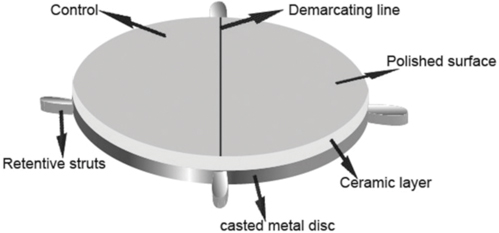
Fig. 1 Porcelain sample with demarcating line in the center.

Fig. 2 Lower retention plates with embedded porcelain samples.
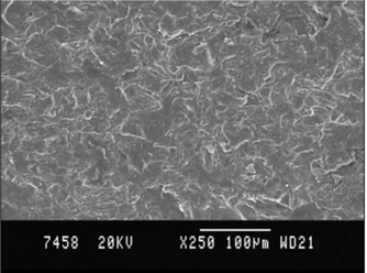
Fig. 3 Scanning electron microscopic photograph of roughened porcelain sample surface (×250).
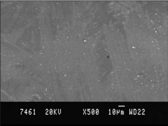
Fig. 4 Scanning electron microscopic photograph of overglazed (Group O) porcelain sample surface (×500).
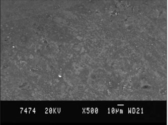
Fig. 5 Scanning electron microscopic photograph of autoglazed (Group A) porcelain sample surface (×500).
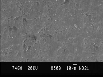
Fig. 6 Scanning electron microscopic photograph of porcelain sample surface polished with Kit S, Shofu (×500).
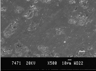
Fig. 7 Scanning electron microscopic photograph of porcelain sample surface polished with Kit D, DFS (×500).
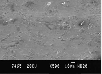
Fig. 8 Scanning electron microscopic photograph of porcelain sample surface polished with Kit E, EVE (×500).
Reference
-
1. Rosenstiel SF, Land MF, Fujimoto J. Contemporary Fixed prosthodontics. 2006. 4th ed. St. Louis: CV Mosby;631.2. Gildenhuys RR, Stallard RE. Comparison of plaque accumulation on metal restorative surfaces. Dent Surv. 1975. 51:56–59.3. Clayton JA, Green E. Roughness of pontic materials and dental plaque. J Prosthet Dent. 1970. 23:407–411.4. Sorensen JA. A rationale for comparison of plaque-retaining properties of crown systems. J Prosthet Dent. 1989. 62:264–269.5. Kaqueler J, Weiss M. Plaque accumulation on dental restorative materials. J Dent Res. 1970. 49:202.6. Keenan MP, Shillingburg HT Jr, Duncanson MG Jr, Wade CK. Effects of cast gold surface finishing on plaque retention. J Prosthet Dent. 1980. 43:168–173.7. Swartz ML, Phillips RW. Comparison of bacterial accumulation on rough and smooth enamel surfaces. J Periodontol. 1957. 28:304–307.8. Wise MD, Dykema RW. The plaque-retaining capacity of four dental materials. J Prosthet Dent. 1975. 33:178–190.9. Henry PJ, Johnston JF, Mitchell DF. Tissue changes beneath fixed partial dentures. J Prosthet Dent. 1966. 16:937–947.10. Quirynen M, Marechal M, Busscher HJ, Weerkamp AH, Darius PL, van Steenberghe D. The influence of surface free energy and surface roughness on early plaque formation. An in vivo study in man. J Clin Periodontol. 1990. 17:138–144.11. Quirynen M, Van der Mei HC, Bollen CM, Van den Bossche LH, Doornbusch GI, van Steenberghe D, Busscher HJ. The influence of surface-free energy on supra- and subgingival plaque microbiology. An in vivo study on implants. J Periodontol. 1994. 65:162–167.12. Quirynen M, Bollen CM. The influence of surface roughness and surface-free energy on supra- and subgingival plaque formation in man. A review of the literature. J Clin Periodontol. 1995. 22:1–14.13. Podshadley AG, harrison JD. Rat connective tissue response to pontic materials. J Prosthet Dent. 1966. 16:110–118.14. Giordano RA 2nd, Campbell S, Pober R. Flexural strength of feldspathic porcelain treated with ion exchange, overglaze, and polishing. J Prosthet Dent. 1994. 71:468–472.15. Williamson RT, Kovarik RE, Mitchell RJ. Effects of grinding, polishing, and overglazing on the flexure strength of a high-leucite feldspathic porcelain. Int J Prosthodont. 1996. 9:30–37.16. al-Hiyasat AS, Saunders WP, Sharkey SW, Smith GM, Gilmour WH. The abrasive effect of glazed, unglazed, and polished porcelain on the wear of human enamel, and the influence of carbonated soft drinks on the rate of wear. Int J Prosthodont. 1997. 10:269–282.17. Monasky GE, Taylor DF. Studies on the wear of porcelain, enamel, and gold. J Prosthet Dent. 1971. 25:299–306.18. Fairhurst CW, Lockwood PE, Ringle RD, Thompson WO. The effect of glaze on porcelain strength. Dent Mater. 1992. 8:203–207.19. Wiley MG. Effects of porcelain on occluding surfaces of restored teeth. J Prosthet Dent. 1989. 61:133–137.20. Jagger DC, Harrison A. An in vitro investigation into the wear effects of unglazed, glazed, and polished porcelain on human enamel. J Prosthet Dent. 1994. 72:320–323.21. Sulik WD, Plekavich EJ. Surface finishing of dental porcelain. J Prosthet Dent. 1981. 46:217–221.22. Klausner LH, Cartwright CB, Charbeneau GT. Polished versus autoglazed porcelain surfaces. J Prosthet Dent. 1982. 47:157–162.23. Scurria MS, Powers JM. Surface roughness of two polished ceramic materials. J Prosthet Dent. 1994. 71:174–177.24. Ward MT, Tate WH, Powers JM. Surface roughness of opalescent porcelains after polishing. Oper Dent. 1995. 20:106–110.25. Patterson CJ, McLundie AC, Stirrups DR, Taylor WG. Efficacy of a porcelain refinishing system in restoring surface finish after grinding with fine and extra-fine diamond burs. J Prosthet Dent. 1992. 68:402–406.26. el-Karaksi AO, Shehab GI, Eskander ME. Effect of reglazing and of polishing on the surface roughness of new ceramic restorations (Hi-ceram). Egypt Dent J. 1993. 39:485–490.27. Chu FC, Frankel N, Smales RJ. Surface roughness and flexural strength of self-glazed, polished, and reglazed In-Ceram/Vitadur Alpha porcelain laminates. Int J Prosthodont. 2000. 13:66–71.
- Full Text Links
-
- Actions
-
Cited
- CITED
-
- Close
- Share
-
- Similar articles
-
- A STUDY ON THE SURFACE ROUGHNESS OF GLAZED PORCELAIN AND POLISHED PORCELAIN
- ANALYSIS OF PORCELAIN SURFACE ROUGHNESS POLISHED BY VARIOUS TECHNIQUE
- The effect of surface finishes on flexural strength, fracture toughness of feldspathic dental porcelain
- AN IN-VITRO WEAR STUDY OF DENTAL PORCELAINS AND HUMAN ENAMEL
- The effect of various polishing systems on surface roughness and phase transformation of monolithic zirconia
