Histomorphometry and stability analysis of early loaded implants with two different surface conditions in beagle dogs
- Affiliations
-
- 1Department of Prosthodontics and Research Institute of Oral Science, College of Dentistry, Kangnung National University, Gangneung, Korea. lila@kangnung.ac.kr
- KMID: 2176333
- DOI: http://doi.org/10.4047/jap.2009.1.1.10
Abstract
- STATEMENT OF PROBLEM: Despite an improved bone reactions of Mg-incorporated implants in the animals, little yet has been carried out by the experimental investigations in functional loading conditions. PURPOSE: This study investigated the clinical and histologic parameters of osseointegrated Mg-incorporated implants in early loading conditions. MATERIAL AND METHODS: A total of 36 solid screw implants (diameter 3.75 mm, length 10 mm) were placed in the mandibles of 6 beagle dogs. Test groups included 18 Mg-incorporated implants. Turned titanium implants served as control. Gold crowns were inserted 4 weeks after implant placement and the dogs were immediately put on a food diet. Implants were observed for 10 weeks after loading. Radiographic assessments and stability tests were performed at the time of fixture installation, 2nd stage surgery, 4 weeks after loading, and 10 weeks after loading. Histological observations and morphometrical measurements were also performed. RESULTS: Of 36 implants, 33 displayed no discernible mobility, corresponding to successful clinical function. There was no statistically significant difference between test implants and controls in marginal bone levels (P = .46) and RFA values. The mean BIC% in the Mg-implants was 54.5 +/- 8.4%. The mean BIC% in the turned implant was 45.3 +/- 12.2%. These differences between the Mg-implant and control implant were statistically significant (P = .005). CONCLUSIONS: The anodized, Mg-incorporated implant demonstrated significantly more bone-to-implant contact (BIC) in early loading conditions. CLINICAL IMPLICATIONS: The results of this study in beagle dogs suggest the possibility of achieving predictable stability of early loaded free-standing dental implants with Mg-incorporated surface.
Keyword
Figure
-
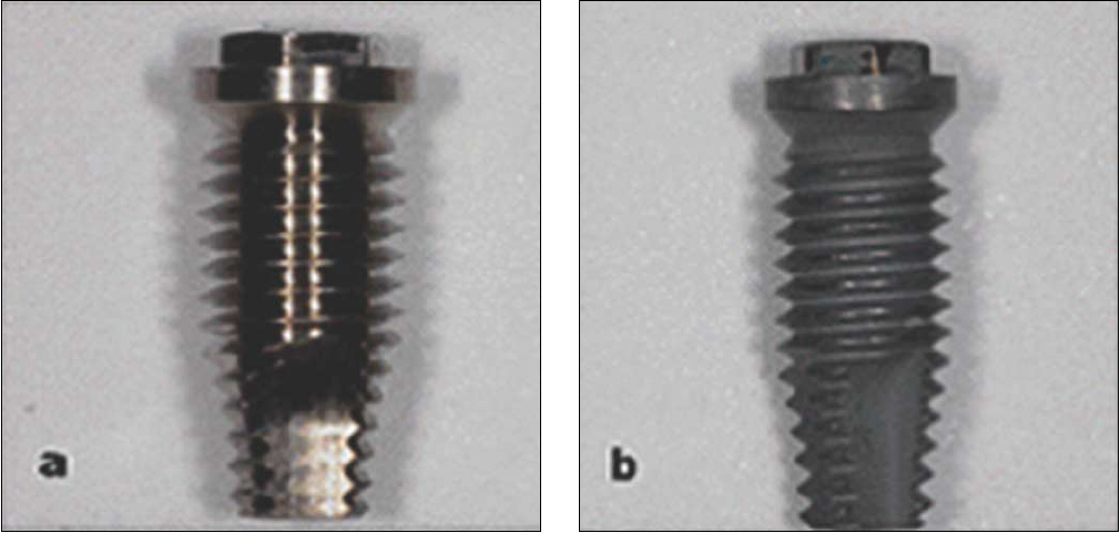
Fig. 1 Control turned implant (a) and test Mg-implant (b).
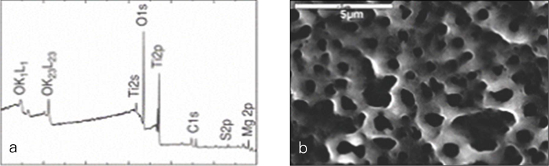
Fig. 2 XPS survey spectra (a) and SEM micrograph of Mg-implant surface (b).
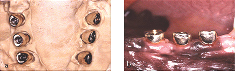
Fig. 3 Delivery of crowns; Crowns in the model (a) and in the mouth (b).

Fig. 4 customized XCP device (a) and periapical radiograph (b).
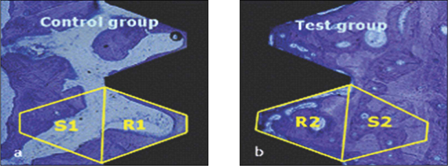
Fig. 5 Bone area within the threads (R1, R2) and their mirror image areas (S1, S2).
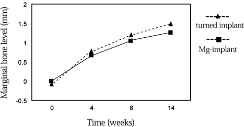
Fig. 6 Marginal bone level change.
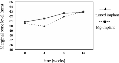
Fig. 7 ISQ value change.

Fig. 8 Bone-implant contact area.
Cited by 1 articles
-
Volumetric quantification of bone-implant contact using micro-computed tomography analysis based on region-based segmentation
Sung-Won Kang, Woo-Jin Lee, Soon-Chul Choi, Sam-Sun Lee, Min-Suk Heo, Kyung-Hoe Huh, Tae-Il Kim, Won-Jin Yi
Imaging Sci Dent. 2015;45(1):7-13. doi: 10.5624/isd.2015.45.1.7.
Reference
-
1. Brånemark PI, Hansson BO, Adell R, Breine U, Lindström J, Hallén O, Ohman A. Osseointegrated implants in the treatment of the edentulous jaw. Experience from a 10-year period. Scand J Plast Reconstr Surg Suppl. 1977. 16:1–132.2. Randow K, Ericsson I, Nilner K, Petersson A, Glantz PO. Immediate functional loading of Brånemark dental implants. An 18-month clinical follow-up study. Clin Oral Implants Res. 1999. 10:8–15.3. Albrektsson T, Zarb G, Worthington P, Eriksson AR. The long-term efficacy of currently used dental implants: a review and proposed criteria of success. Int J Oral Maxillofac Implants. 1986. 1:11–25.4. Albrektsson T, Sennerby L. State of the art in oral implants. J Clin Periodontol. 1991. 18:474–481.5. Brunski JB. Avoid pitfalls of overloading and micromotion of intraosseous implants. Dent Implantol Update. 1993. 4:77–81.6. Szmukler-Moncler S, Salama H, Reingewirtz Y, Dubruille JH. Timing of loading and effect of micromotion on bone-dental implant interface: review of experimental literature. J Biomed Mater Res. 1998. 43:192–203.7. Gomes A, Lozada JL, Caplanis N, Kleinman A. Immediate loading of a single hydroxyapatite-coated threaded root form implant: a clinical report. J Oral Implantol. 1998. 24:159–166.8. Piattelli A, Corigliano M, Scarano A, Quaranta M. Bone reactions to early occlusal loading of two-stage titanium plasma-sprayed implants: a pilot study in monkeys. Int J Periodontics Restorative Dent. 1997. 17:162–169.9. Piattelli A, Corigliano M, Scarano A. Microscopical observations of the osseous responses in early loaded human titanium implants: a report of two cases. Biomaterials. 1996. 17:1333–1337.10. Lefkove MD, Beals RP. Immediate loading of cylinder implants with overdentures in the mandibular symphysis: the titanium plasma-sprayed screw technique. J Oral Implantol. 1990. 16:265–271.11. Misch CE. Implant design considerations for the posterior regions of the mouth. Implant Dent. 1999. 8:376–386.12. Hench LL, Paschall HA. Direct chemical bond of bioactive glass-ceramic materials to bone and muscle. J Biomed Mater Res. 1973. 7:25–42.13. Ishizawa H, Ogino M. Formation and characterization of anodic titanium oxide films containing Ca and P. J Biomed Mater Res. 1995. 29:65–72.14. Sul YT, Johansson CB, Kang Y, Jeon DG, Albrektsson T. Bone reactions to oxidized titanium implants with electrochemical anion sulphuric acid and phosphoric acid incorporation. Clin Implant Dent Relat Res. 2002. 4:78–87.15. Sul YT, Byon ES, Jeong Y. Biomechanical measurements of calcium-incorporated oxidized implants in rabbit bone: effect of calcium surface chemistry of a novel implant. Clin Implant Dent Relat Res. 2004. 6:101–110.16. Sul YT, Johansson C, Byon E, Albrektsson T. The bone response of oxidized bioactive and non-bioactive titanium implants. Biomaterials. 2005. 26:6720–6730.17. Martin JY, Schwartz Z, Hummert TW, Schraub DM, Simpson J, Lankford J Jr, Dean DD, Cochran DL, Boyan BD. Effect of titanium surface roughness on proliferation, differentiation, and protein synthesis of human osteoblast-like cells (MG63). J Biomed Mater Res. 1995. 29:389–401.18. Zreiqat H, Howlett CR, Zannettino A, Evans P, Schulze-Tanzil G, Knabe C, Shakibaei M. Mechanisms of magnesium-stimulated adhesion of osteoblastic cells to commonly used orthopaedic implants. J Biomed Mater Res. 2002. 62:175–184.19. Lapidos KA, Woodhouse EC, Kohn EC, Masiero L. Mg(++)-induced endothelial cell migration: substratum selectivity and receptor-involvement. Angiogenesis. 2001. 4:21–28.20. Donath K. . Preparation of histologic sections by cutting-grinding technique for hard tissue and other materials not suitable to be sectioned by routine methods. 1993. Norderstedt: EXAKT-Kulzer-Publication;1–6.21. Akagawa Y, Ichikawa Y, Nikai H, Tsuru H. Interface histology of unloaded and early loaded partially stabilized zirconia endosseous implant in initial bone healing. J Prosthet Dent. 1993. 69:599–604.22. Sagara M, Akagawa Y, Nikai H, Tsuru H. The effects of early occlusal loading on one-stage titanium alloy implants in beagle dogs: a pilot study. J Prosthet Dent. 1993. 69:281–288.23. Meredith N, Shagaldi F, Alleyne D, Sennerby L, Cawley P. The application of resonance frequency measurements to study the stability of titanium implants during healing in the rabbit tibia. Clin Oral Implants Res. 1997. 8:234–243.24. Friberg B, Sennerby L, Linden B, Gröndahl K, Lekholm U. Stability measurements of one-stage Brånemark implants during healing in mandibles. A clinical resonance frequency analysis study. Int J Oral Maxillofac Surg. 1999. 28:266–272.25. O'Sullivan D, Sennerby L, Meredith N. Measurements comparing the initial stability of five designs of dental implants: a human cadaver study. Clin Implant Dent Relat Res. 2000. 2:85–92.26. Larsson C, Thomsen P, Lausmaa J, Rodahl M, Kasemo B, Ericson LE. Bone response to surface modified titanium implants: studies on electropolished implants with different oxide thicknesses and morphology. Biomaterials. 1994. 15:1062–1074.27. de Lange GL, Donath K. Interface between bone tissue and implants of solid hydroxyapatite or hydroxyapatite-coated titanium implants. Biomaterials. 1989. 10:121–125.28. Buser D, Nydegger T, Hirt HP, Cochran DL, Nolte LP. Removal torque values of titanium implants in the maxilla of miniature pigs. Int J Oral Maxillofac Implants. 1998. 13:611–619.29. Davies JE. In vitro modeling of the bone/implant interface. Anat Rec. 1996. 245:426–445.30. Wong M, Eulenberger J, Schenk R, Hunziker E. Effect of surface topology on the osseointegration of implant materials in trabecular bone. J Biomed Mater Res. 1995. 29:1567–1575.31. Cochran DL. The scientific basis for and clinical experiences with Straumann implants including the ITI Dental Implant System: a consensus report. Clin Oral Implants Res. 2000. 11:33–58.32. Buser D, Schenk RK, Steinemann S, Fiorellini JP, Fox CH, Stich H. Influence of surface characteristics on bone integration of titanium implants. A histomorphometric study in miniature pigs. J Biomed Mater Res. 1991. 25:889–902.33. Larsson C, Thomsen P, Lausmaa J, Rodahl M, Kasemo B, Ericson LE. Bone response to surface modified titanium implants: studies on electropolished implants with different oxide thicknesses and morphology. Biomaterials. 1994. 15:1062–1074.34. Chen G, Wen X, Zhang N. Corrosion resistance and ion dissolution of titanium with different surface microroughness. Biomed Mater Eng. 1998. 8:61–74.35. McAlarney ME, Oshiro MA, McAlarney CV. Effects of titanium dioxide passive film crystal structure, thickness, and crystallinity on C3 adsorption. Int J Oral Maxillofac Implants. 1996. 11:73–80.36. Fujibayashi S, Nakamura T, Nishiguchi S, Tamura J, Uchida M, Kim HM, Kokubo T. Bioactive titanium: effect of sodium removal on the bone-bonding ability of bioactive titanium prepared by alkali and heat treatment. J Biomed Mater Res. 2001. 56:562–570.37. Li J. Behaviour of titanium and titania-based ceramics in vitro and in vivo. Biomaterials. 1993. 14:229–232.
- Full Text Links
-
- Actions
-
Cited
- CITED
-
- Close
- Share
-
- Similar articles
-
- Bone response of three different surface implants : Histomorphometric, perio test value and resonance frequency analysis in beagle dogs
- Bone response of three different surface implants: histomorphometric and resonance frequency analysis in dogs
- Comparision of Osseointegration Depending on Surface Treatment
- Effects of drilling process in stability of micro-implants used for the orthodontic anchorage
- Comparion of stability in titanium implants with different surface topographies in dogs
