J Korean Acad Conserv Dent.
2009 May;34(3):232-239. 10.5395/JKACD.2009.34.3.232.
Effects of anticurvature filing on danger zone width in curved root canals
- Affiliations
-
- 1Department of Conservative Dentistry, College of Dentistry, Yonsei University, Korea. operatys16@yuhs.ac
- 2Oral Science Research Center, College of Dentistry, Yonsei University, Korea.
- KMID: 2176149
- DOI: http://doi.org/10.5395/JKACD.2009.34.3.232
Abstract
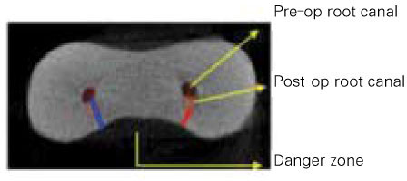
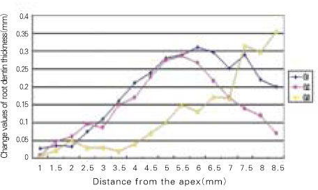
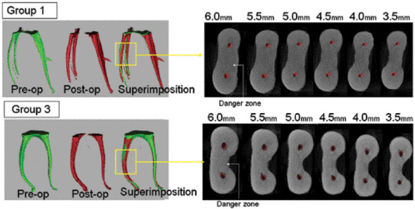
- The aim of this study was to compare the effects of anticurvature filing with stainless steel k-file versus nickel-titanium ProFile in the shaping of mesial root canals of extracted mandibular molars. A total of 60 canals from 30 mesial roots of mandibular molar teeth were randomly assigned to three groups with n=20 each. They were prepared with different instruments and methods: The first group with stainless steel k-file and circumferential filing, the second with precurved stainless steel k-file and anticurvature filing and the third with ProFile (.06 taper) and anticurvature filing. Using a micro-computed tomography system (skyscan-1076, SKYSCAN, Antwerpen, Belgium), pre-and post-operative specimens were scanned. Subsequently, canal images were superimposed and changes in root dentin thickness were measured at distal side (danger zone) of the canal. The data was analyzed using a one-way ANOVA and the comparison of means was conducted using a post hoc multiple comparison Tukey test. There were significant differences in the change of root dentin thickness at the 7.5~8.5mm level between group 1 and 2, 3.5~6mm level between group 1 and 3 and 3.5~6mm level between group 2 and 3(n=20, P<0.05).
Figure
Reference
-
1. Schilder H. Clean and shaping the root canal. Dent Clin North Am. 1974. 18:269–296.2. Bishop K, Dummer PM. A comparison of stainless steel Flexofiles and nickel-titanium NiTiFlex files during the shaping of simulated canals. Int Endod J. 1997. 30:25–34.
Article3. Schneider SW. A comparison of the canal preparations in straight and curved root canals. Oral surg. 1971. 32:271–275.4. Weine F, Kelly RF, Lio PJ. The effect of proparation procedures on original canal shape and on apical foramen shape. J Endod. 1975. 1:255–262.
Article5. Meister F Jr, Lommel TJ, Gerstein H. Endodontic perforations which resulted in alveolar bone loss. Report of five cases. Oral Surg Oral Med Oral Pathol. 1979. 47(5):463–470.6. Abou-Rass M, Frank AL, Glick DH. The anticurvature filing method to prepare the curved root canal. J Am Dent Assoc. 1980. 101(5):792–794.
Article7. Goerig AC, Michelich RJ, Schultz H. Instrumentation of root canals in molar using step-down technique. J Endod. 1982. 8:550–554.8. Walia HM, Brantley WA, Gerstein H. An initial investigation of the bending and torsional properties of Nitinol root canal files. J Endod. 1988. 14:346–351.
Article9. Glossen CR, Haller RH, Dove SB, del Rio CE. A comparison of root canal preparation using Ni-Ti hand, Ni-Ti engine-driven, and K-Flex endodontic instruments. J Endod. 1995. 21(3):146–151.10. Thompson SA, Dummer PM. Shaping ability of Hero 642 rotary nickel-titanium instruments in simulated root canals: Part 1. Int Endod J. 2000. 33:248–254.
Article11. Tachibana H, Matsumoto K. Application of x-ray computed tomography in endodontics. Endod Dent Traumatol. 1990. 6(1):16–20.12. Nielsen RB, Alyassin AM, et al. Microcomputed tomography: An advanced system for detailed endodontic researc. J Endod. 1995. 21:561–568.13. Gambill JM, Alder M, del Rio CE. Comparison of nikeltitanium and stainless steel hand file instrumentation using computed tomography. J Endod. 1996. 22:369–375.
Article14. Rhodes JS, Ford TR, et al. Micro-computed tomography: a new tool for experimental endodontology. Int Endod J. 1999. 32:165–170.
Article15. Gluskin AH, Brown DC. A reconstructed computerized tomographic comparison of Ni-Ti rotary GT files versus traditional instruments in canals shaped by novice operators. Int Endod J. 2001. 34(6):476–484.
Article16. Schäfer E. Shaping ability of Hero 642 rotary nickel-titanium instruments and stainless steel hand K-Flexofiles in simulated curved root canals. Oral Surg Oral Med Oral Pathol Oral Radiol Endod. 2001. 92:215–220.
Article17. Esposito PT, Cunningham CJ. A comparison of canal preparation with nickel-titanium and stainless steel instrumentation. J Endod. 1995. 21:173–176.
Article18. Peters OA, Laib A, Ruegsegger P, Barbakow F. Threedimensional analysis of root canal geometry by high resolution computed tomography. J Dent Res. 2000. 79(6):1405–1409.
Article19. Bergmans L, Van Cleynenbreugel J, Wevers M. A methodology for quantitative evaluation using microcomputed tomography. Int Endod J. 2001. 34(5):390–398.
Article20. Lim SS, Stock CJ. The risk of perforation in the curved canal : anticurvature filing compared with the step-back technique. Int Endod J. 1987. 20(1):33–39.
Article21. Kessler JR, Peters DD, Lorton L. Comparison of the relative risk of molar root perforation using various endodontic instrumentation techniques. J Endod. 1983. 9:439–447.22. Kosa DA, Marshall G, Baumgartner JC. An analysis of canal centering using mechanical instrumentation techniques. J Endod. 1999. 25:441–445.
Article
- Full Text Links
- Actions
-
Cited
- CITED
-
- Close
- Share
- Similar articles
-
- Shaping ability of four rotary nickel-titanium instruments to prepare root canal at danger zone
- Effect of anticurvature filing method on preparation of the curved root canal using ProFile
- Endodontic treatment of a C-shaped mandibular second premolar with four root canals and three apical foramina: a case report
- Assessment of Root and Root Canal Morphology of Human Primary Molars using CBCT
- Impact of root canal curvature and instrument type on the amount of extruded debris during retreatment


