Ductal Carcinoma Arising from Ectopic Breast Tissue Following Microcalcification Observed on Screening Mammography: A Case Report and Review of the Literature
- Affiliations
-
- 1Department of Surgery, Breast Cancer Center, Kyungpook National University Medical Center, Daegu, Korea. phy123@knu.ac.kr
- 2Department of Radiology, Breast Cancer Center, Kyungpook National University Medical Center, Daegu, Korea.
- 3Department of Pathology, Breast Cancer Center, Kyungpook National University Medical Center, Daegu, Korea.
- 4Dr. Lim's Breast Clinic, Daegu, Korea.
- KMID: 2176132
- DOI: http://doi.org/10.4048/jbc.2014.17.4.393
Abstract
- Ectopic breast tissue can occur anywhere along the incompletely regressed mammary ridge. Among the various types of breast choristoma, ectopic breast tissue, which has only glandular tissue without a nipple or areola, is most commonly detected in axillary areas. However, ectopic breast cancer is often not detected until significant clinical symptoms have been revealed, or diagnosis is delayed. Furthermore, an examination of ectopic breast tissue tends to be omitted from a screening mammography. Especially, the microcalcifications of ectopic breast tissue are difficult to delineate on mammography. Herein, the authors report a case of ectopic breast carcinoma that showed clustered microcalcifications on screening mammography, and discuss the interpretation and implications of microcalcification in ectopic breast tissue.
Keyword
MeSH Terms
Figure
-

Figure 1 Mammographic findings of ectopic breast cancer. (A) Aberrant breast tissue with clustered microcalcifications (dot circle) is detected on right axilla. (B) Magnified mammographic image of ectopic breast tissue with microcalcifications. A clustered microcalcification is well observed on ectopic breast tissue. (C) Mammographic findings of removed breast tissue. Multifocal microcalcification is apparent in two separately removed ectopic breast tissue specimens.
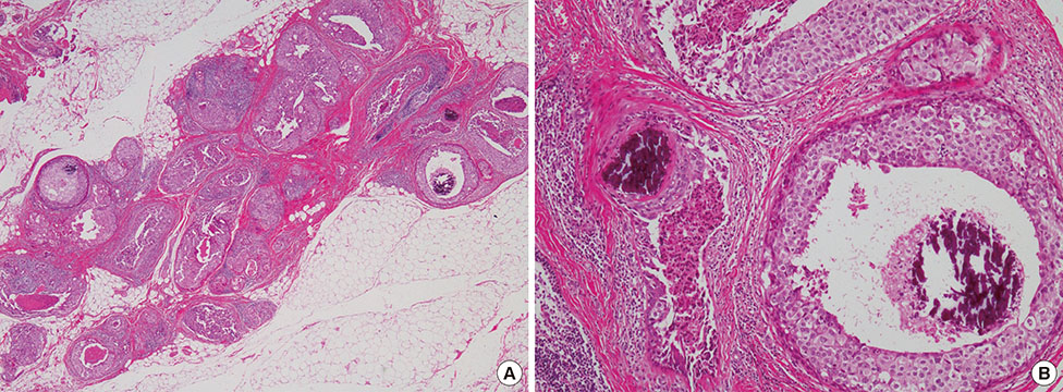
Figure 2 Histopathologic findings of ectopic breast carcinoma. (A) Ductal epithelial proliferative lesion with multiple foci of necrosis and microcalcification is noted (H&E stain, ×20). (B) The tumor cells show moderate to high degree of nuclear atypia characteristic of high-grade ductal carcinoma in situ. Microcalcification is well observed in the tumor cell nests (H&E stain, ×100).
Reference
-
1. Kajava Y. The proportions of supernumerary nipples in the Finnish population. Duodecim. 1915; 31:143–170.2. Marshall MB, Moynihan JJ, Frost A, Evans SR. Ectopic breast cancer: case report and literature review. Surg Oncol. 1994; 3:295–304.
Article3. Francone E, Nathan MJ, Murelli F, Bruno MS, Traverso E, Friedman D. Ectopic breast cancer: case report and review of the literature. Aesthetic Plast Surg. 2013; 37:746–749.
Article4. Stomper PC, Geradts J, Edge SB, Levine EG. Mammographic predictors of the presence and size of invasive carcinomas associated with malignant microcalcification lesions without a mass. AJR Am J Roentgenol. 2003; 181:1679–1684.
Article5. Venkatesan A, Chu P, Kerlikowske K, Sickles EA, Smith-Bindman R. Positive predictive value of specific mammographic findings according to reader and patient variables. Radiology. 2009; 250:648–657.
Article6. Singer C, Blankstein E, Koenigsberg T, Mercado C, Pile-Spellman E, Smith SJ. Mammographic appearance of axillary lymph node calcification in patients with metastatic ovarian carcinoma. AJR Am J Roentgenol. 2001; 176:1437–1440.
Article7. Burdeny DA, Reed MH, Ferguson CA. Calcification of axillary lymph nodes following BCG vaccination. Can Assoc Radiol J. 1989; 40:92–93.8. Chen SW, Bennett G, Price J. Axillary lymph node calcification due to metastatic papillary carcinoma. Australas Radiol. 1998; 42:241–243.
Article9. Amsler E, Sigal-Zafrani B, Marinho E, Aractingi S. Ectopic breast cancer of the axilla. Ann Dermatol Venereol. 2002; 129:1389–1391.10. Yerra L, Karnad AB, Votaw ML. Primary breast cancer in aberrant breast tissue in the axilla. South Med J. 1997; 90:661–662.
Article11. Nihon-Yanagi Y, Ueda T, Kameda N, Okazumi S. A case of ectopic breast cancer with a literature review. Surg Oncol. 2011; 20:35–42.
Article12. Das DK, Gupta SK, Mathew SV, Sheikh ZA, al-Rabah NA. Fine needle aspiration cytologic diagnosis of axillary accessory breast tissue, including its physiologic changes and pathologic lesions. Acta Cytol. 1994; 38:130–135.13. Bakker JR, Sataloff DM, Haupt HM. Breast cancer presenting in aberrant axillary breast tissue. Community Oncol. 2005; 2:117–122.
Article14. Kim TW, Kang SW, Park JY, Ko SS, Hur MH, Lee HK, et al. Invasive ductal carcinoma arising from axillary accessory breast. J Korean Breast Cancer Soc. 2004; 7:306–310.
Article15. Alghamdi H, Abdelhadi M. Accessory breasts: when to excise. Breast J. 2005; 11:155–157.
Article
- Full Text Links
-
- Actions
-
Cited
- CITED
-
- Close
- Share
-
- Similar articles
-
- Concurrent Invasive Carcinoma and Fibroadenoma Arising from Bilateral Ectopic Breast Tissue in the Chest Wall: A Case Report and Literature Review
- Invasive Ductal Carcinoma Arising within a Mammary Hamartoma: Case Report
- An Unusual Presentation of Extensive Ductal Carcinoma in Situ Accompanying Invasive Ductal Carcinoma on MRI: A Case Report
- Ductal Carcinoma in Situ: US Finding and Its Usefulness
- Primary Invasive Ductal Carcinoma Arising in Axillary Accessory Breast: A Case Report
