Effect of restoration type on the stress distribution of endodontically treated maxillary premolars; Three-dimensional finite element study
- Affiliations
-
- 1Department of Conservative dentistry, School of Dentistry, Pusan National University, Korea. jeongkil@pusan.ac.kr
- 2Department of Mechanical design engineering, College of Engineering, Pusan National University, Korea.
- KMID: 2176038
- DOI: http://doi.org/10.5395/JKACD.2009.34.1.008
Abstract
- The purpose of this study was to investigate the effects of four restorative materials under various occlusal loading conditions on the stress distribution at the CEJ of buccal, palatal surface and central groove of occlusal surface of endodontically treated maxillary second premolar, using a 3D finte element analysis. A 3D finite element model of human maxillary second premolar was endodontically treated. After endodontic treatment, access cavity was filled with Amalgam, resin, ceramic or gold of different mechanical properties. A static 500N forces were applied at the buccal (Load-1) and palatal cusp (Load-2) and a static 170N forces were applied at the mesial marginal ridge and palatal cusp simultaneously as centric occlusion (Load-3). Under 3-type Loading condition, the value of tensile stress was analyzed after 4-type restoration at the CEJ of buccal and palatal surface and central groove of occlusal surface. Excessive high tensile stresses were observed along the palatal CEJ in Load-1 case and buccal CEJ in Load-2 in all of the restorations. There was no difference in magnitude of stress in relation to the type of restorations. Heavy tensile stress concentrations were observed around the loading point and along the central groove of occlusal surface in all of the restorations. There was slight difference in magnitude of stress between different types of restorations. High tensile stress concentrations around the loading points were observed and there was no difference in magnitude of stress between different types of restorations in Load-3.
Keyword
Figure
-

Figure 1 Access cavity restoration (light brown; GI base, dark brown: restorative material).
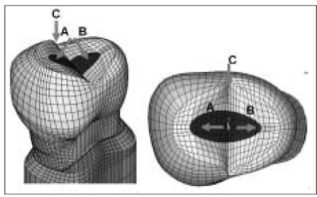
Figure 2 Three load conditions of 3D FE model. Load-1: loading at A point (500 N) Load-2: loading at B point (500 N) Load-3: simultaneous loading at B point (100 N) and C point (70 N)
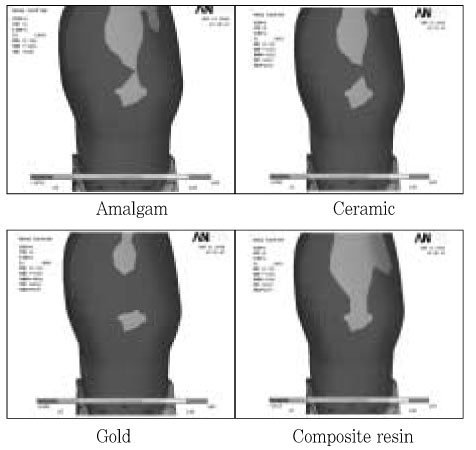
Figure 3 The buccal view of maximum principal stress distribution under Load-1.

Figure 4 The maximum principal stress distribution along the buccal CEJ under Load-1.

Figure 5 The palatal view of maximum principal stress distribution under Load-1.
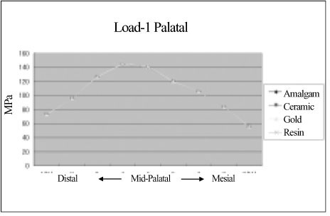
Figure 6 The maximum principal stress of CEJ of palatal surface under Load-1.
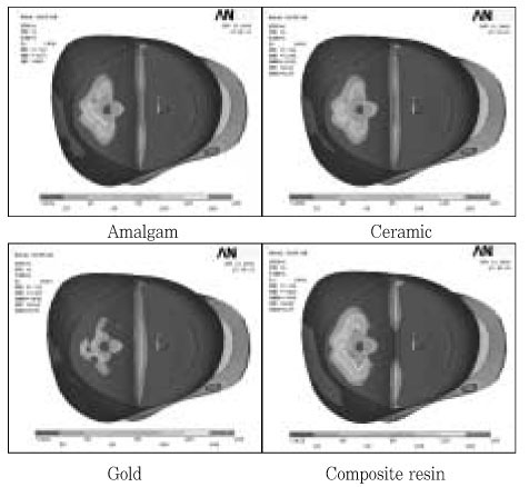
Figure 7 Different stress patterns of occlusal surface under Load-1.
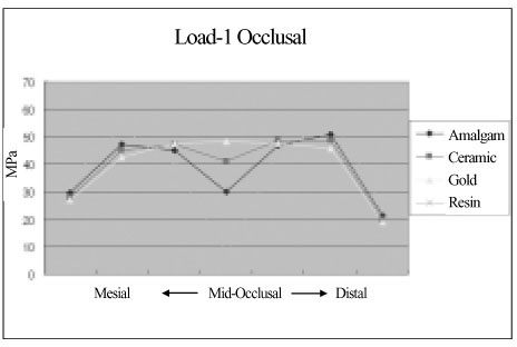
Figure 8 The maximum principal stress distribution along the central groove of occlusal surface under Load-1.
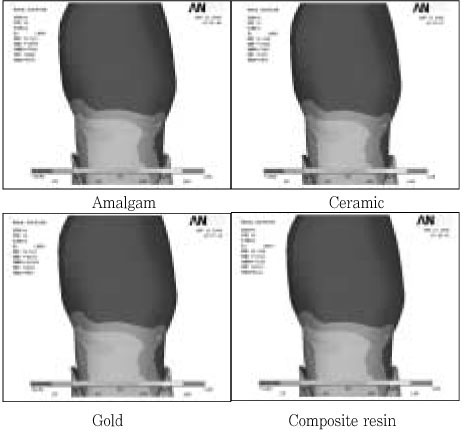
Figure 9 The buccal view of maximum principal stress distribution under Load-2.

Figure 10 The maximum principal stress distribution along the CEJ of buccal surface under Load-2.
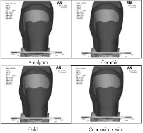
Figure 11 The palatal view of maximum principal stress distribution under Load-2.

Figure 12 The maximum principal stress distribution along the palatal CEJ under Load-2.

Figure 13 The occlusal view of maximum principal stress distribution under Load-2.
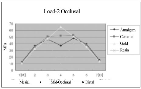
Figure 14 The maximum principal stress analysis stress distribution along central groove of occlusal groove under Load-2.
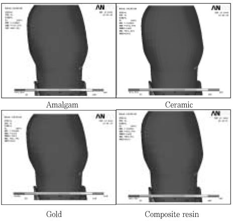
Figure 15 The Buccal view of maximum principal stress distribution under Load-3.

Figure 16 The maximum principal stress distribution along the Buccal CEJ under Load-3.
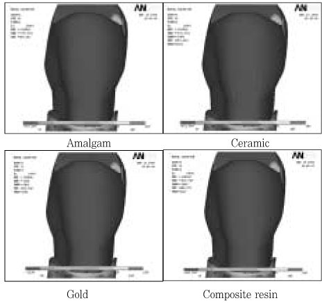
Figure 17 The palatal view of maximum principal stress distribution under Load-3,
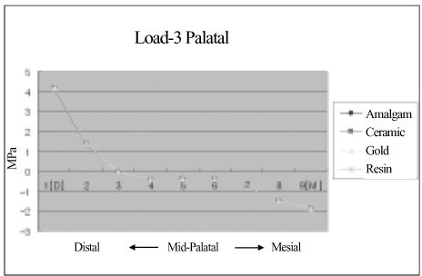
Figure 18 The maximum principal stress distribution along the palatal CEJ under Load-3.
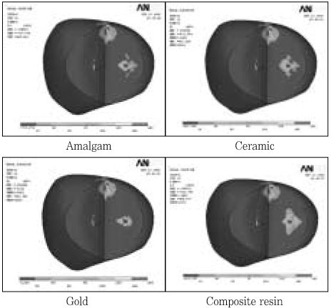
Figure 19 The occlusal view of maximum principal stress distribution under Load-3.
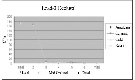
Figure 20 The maximum principal stress distribution along the central groove of occlusal surface under Load-3.
Reference
-
1. Hannig C, Westphal C, Becker K, Attin T. Fracture resistance of endodontically treated maxillary premolars restored with CAD/CAM ceramic inlays. J Prosthet Dent. 2005. 94:342–349.
Article2. Panitvisai P, Messer HH. Cuspal deflection in molars in relation to endodontic and restorative procedures. J Endod. 1995. 21:57–61.
Article3. Reeh ES, Messer HH, Douglas WH. Reduction in tooth stiffness as a result of endodontic and restorative procedures. J Endod. 1989. 15:512–516.
Article4. Trope M, Langer I, Maltz D, Tronstad L. Resistance to fracture of restored endodontically treated premolars. Endod Dent Traumatol. 1986. 2:35–38.
Article5. Reeh ES, Douglas WH, Messer HH. Stiffness of endodontically-treated teeth related to restoration technique. J Dent Res. 1989. 68:1540–1544.
Article6. Sorensen JA, Martinoff JT. Intracoronal reinforcement and coronal coverage: a study of endodontically treated teeth. J Prosthet Dent. 1984. 51:780–784.
Article7. Sedgley CM, Messer HH. Are endodontically treated teeth more brittle? J Endod. 1992. 18:332–335.
Article8. Hofmann N, Just N, Haller B, Hugo B, Klaiber B. The effect of glass ionomer cement or composite resin bases on restoration of cuspal stiffness of endodontically treated premolars in vitro. Clin Oral Investig. 1998. 2:77–83.
Article9. Hernandez R, Bader S, Boston D, Trope M. Resistance to fracture of endodontically treated premolars restored with new generation dentine bonding systems. Int Endod J. 1994. 27:281–284.
Article10. Santos MJ, Bezerra RB. Fracture Resistance of Maxillary Premolars Restored with Direct and Indirect Adhesive Techniques. J Can Dent Assoc. 2005. 71:585.11. Hansen EK. In vivo cusp fracture of endodontically treated premolars restored with MOD amalgam or MOD resin fillings. Dent Mater. 1988. 4:169–173.
Article12. Katona TR, Winkler MM. Stress analysis of a bulk-filled Class V light-cured composite restoration. J Dent Res. 1994. 73:1470–1477.
Article13. Geramy A, Sharafoddin F. Abfraction: 3D analysis by means of the finite element method. Quintessence Int. 2003. 34:526–533.14. Lindehe J, Karring T. Schluger S, Yuodelis R, Page RC, Johnson RH, editors. The anatomy of the periodontium. Textbook of Clinical Periodontology. 1989. 2nd edition. Munksgaard: Copenhagen;19–69.15. Schroeder HE, Page RC. Schluger S, Yuodelis R, Page RC, Johnson RH. The normal periodontium. Periodontal Diseases. 1990. 2nd edition. Lea & Fabiger: Philadelphia;3–52.16. Couegnat G, Fok SL, Cooper JE, Qualtrough AJ. Structural optimization of dental restorations using the principle of adaptive growth. Dent Mater. 2006. 22:3–12.
Article17. Suansuwan S, Swain M. New approach for evaluating metal-porcelain interfacial bonding. Int J Prosthodont. 1999. 12:547–552.18. Ichim I, Schmidlin PR, Kieser JA, Swain MV. Mechanical evaluation of cervical glass-ionomer restorations: 3D finite element study. J Dent. 2007. 35:28–35.
Article19. Widmalm SE, Ericsson SG. Maximal bite force with centric and eccentric load. J Oral Rehabil. 1982. 9:445–450.
Article20. Litonjua LA, Andreana S, Patra AK, Cohen RE. An assessment of stress analyses in the theory of abfraction. Biomed Mater Eng. 2004. 14:311–321.21. Lee HM, Hur B, Kim HC, Woo SG, Kim KH, Son K, Park JK. Effects of occlusal load on the cervical stress distribution : A three-dimensional finite element study. J Korean Acad Conserv Dent. 2006. 31:427–436.
Article22. Lewinstein I, Grajower R. Root dentin hardness of endodontically treated teeth. J Endod. 1981. 7:421–422.
Article23. Yettram AL, Wright KW, Pickard HM. Finite element stress analysis of the crowns of normal and restored teeth. J Dent Res. 1976. 55:1004–1011.
Article24. Park JK, Hur B, Kim SK. Stress distribution of class V composite resin restorations: A three-dimensional finite element study. J Korean Acad Conserv Dent. 2008. 33:36–46.
Article25. Park JK, Hur B, Kim SK. The influence of combining composite resins with different elastic modulus on the stress distribution of class V restoration : A three-dimensional finite element study. J Korean Acad Conserv Dent. 2008. 33:184–197.
Article26. Kuroe T, Itoh H, Caputo AA, Nakahara H. Potential for load-induced cervical stress concentration as a function of periodontal support. J Esthet Dent. 1999. 11:215–222.
Article27. Milicich G, Rainey JT. Clinical presentations of stress distribution in teeth and the significance in operative dentistry. Pract Periodontics Aesthet Dent. 2000. 12:695–700.28. Ortega VL, Pegoraro LF, Conti PC, do Valle AL, Bonfante G. evaluation of fracture resistance of endodontically treated maxillary premolars, restored with ceromer or heat-pressed ceramic inlays and fixed with dual-resin cements. J Oral Rehabil. 2004. 31:393–397.
Article30. Khers SC, Carpenter CW, Vetter JD, Staley RN. Anatomy of cusps of posterior teeth and their fracture potential. J Prosthet Dent. 1990. 64:139–147.
Article
- Full Text Links
-
- Actions
-
Cited
- CITED
-
- Close
- Share
-
- Similar articles
-
- Stress distribution of endodontically treated maxillary second premolars restored with different methods: Three-dimensional finite element analysis
- A three-dimensional Finite element analysis for initial stress of maxillary incisors during activation of upper utility arch wire
- The effect of restorative materials on the stress distribution of class V composite resin restorations: a 3D finite element investigation
- Critical evaluation of fracture strength testing for endodontically treated teeth: a finite element analysis study
- A finite element analysis on the effect of the reverse headgear to the maxillary complex
