Effect of cavity shape, bond quality and volume on dentin bond strength
- Affiliations
-
- 1Department of Conservative Dentistry, School of Dentistry, Seoul National University, Korea. chobh@snu.ac.kr
- 2Dental Research Institute, School of Dentistry, Seoul National University, Korea.
- 3Department of Orthodontics, School of Dentistry, Seoul National University, Korea.
- 4Department of Dental Biomaterials, School of Dentistry, Seoul National University, Korea.
- KMID: 2175749
- DOI: http://doi.org/10.5395/JKACD.2005.30.6.450
Abstract
- The aim of this study was to evaluate the effect of cavity shape, bond quality of bonding agent and volume of resin composite on shrinkage stress developed at the cavity floor. This was done by measuring the shear bond strength with respect to iris materials (cavity shape; adhesive-coated dentin as a high C-factor and Teflon-coated metal as a low C-factor), bonding agents (bond quality; Scotchbond(TM) Multi-purpose and Xeno(R)III) and iris hole diameters (volume; 1 mm or 3 mm in diameter x 1.5 mm in thickness). Ninety-six molars were randomly divided into 8 groups (2 x 2 x 2 experimental setup). In order to simulate a Class I cavity, shear bond strength was measured on the flat occlusal dentin surface with irises. The iris hole was filled with Z250 restorative resin composite in a bulk-filling manner. The data was analyzed using three-way ANOVA and the Tukey test. Fracture mode analysis was also done. When the cavity had high C-factor, good bond quality and large volume, the bond strength decreased significantly. The volume of resin composite restricted within the well-bonded cavity walls is also be suggested to be included in the concept of C-factor, as well as the cavity shape and bond quality. Since the bond quality and volume can exaggerate the effect of cavity shape on the shrinkage stress developed at the resin-dentin bond, resin composites must be filled in a method, which minimizes the volume that can increase the C-factor.
Keyword
Figure
-

Figure 1 Schematic illustration of the experimental set-up. The metal and dentin irises having a hole of 1 mm or 3 mm in diameter and 1.5 mm in thickness were prepared. The internal wall of the metal iris was coated with Teflon and that of the dentin iris was treated with the assigned bonding agent. The irises were put on the flat occlusal dentin surface, which was also treated with the same bonding agent, and resin composite was filled into the hole. The shear bond strength was measured following the chisel-on-iris method.
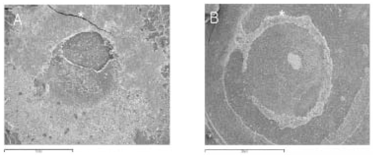
Figure 2 Scanning electron micrograph of the failure surfaces. A. Mixed failure. Fracture fragments of the adhesive layer were observed simultaneously with the detached plain surface at the top of the hybrid layer. The specimen was tested with the dentin iris having a 1 mm hole and SBMP (× 20). B. Typical failure at the top of the hybrid layer observed in the specimen tested with the metal iris having a 3 mm iris hole and Xeno III (magnification: × 20). *: the direction from which the load was applied.
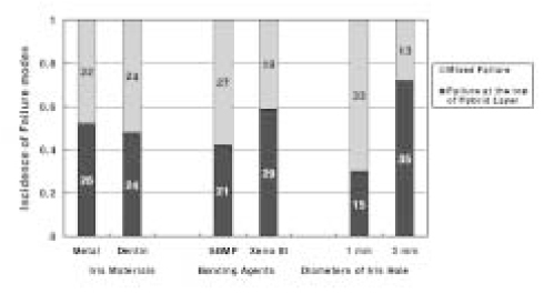
Figure 3 Incidence of failure modes with respect to iris materials (cavity shape), bonding agents (bond quality) and diameters of iris hole (volume). The numbers in the bars are those of the specimens showing 'failure at the top of the hybrid layer' or 'mixed failure'.
Reference
-
1. Feilzer AJ, de GEE AF, Davidson CL. Setting stress in composite resin in relation to configuration of the restoration. J Dent Res. 1987. 66:1636–1639.
Article2. Davidson CL, de Gee AJ, Feilzer A. The competition between the composite-dentin bond strength and the polymerization contraction stress. J Dent Res. 1984. 63:1396–1399.
Article3. Armstrong SR, Keller JC, Boyer DB. The influence of water storage and C-factor on the dentin-resin composite microtensile bond strength and debond pathway utilizing a filled and unfilled adhesive resin. Dent Mater. 2001. 17:268–276.
Article4. Nikaido T, Kunzelmann KH, Chen H, Ogata M, Harada N, Yamaguchi S, Cox CF, Hickel R, Tagami J. Evaluation of thermal cycling and mechanical loading on bond strength of a self-etching primer system to dentin. Dent Mater. 2002. 18:269–275.
Article5. Nikolaenko SA, Lohbauer U, Roggendorf M, Petschelt A, Dasch W, Frankenberger R. Influence of c-factor and layering technique on microtensile bond strength to dentin. Dent Mater. 2004. 20:579–585.
Article6. Uno S, Shimokobe H. Contraction stress and marginal adaptation of composite restorations in dentinal cavity. Dent Mater J. 1994. 13:19–24.
Article7. Versluis A, Tantbirojn D, Douglas WH. Do dental composites always shrink toward the light? J Dent Res. 1998. 77:1435–1445.
Article8. Asmussen E, Peutzfeldt A. Direction of shrinkage of light-curing resin composites. Acta Odontol Scand. 1999. 57:310–315.
Article9. Cho BH, Dickens SH, Bae JH, Chang CG, Son HH, Um CM. Effect of interfacial bond quality on the direction of polymerization shrinkage flow in resin composite restorations. Oper Dent. 2002. 27:297–304.10. Dickens SH, Cho BH. Interpretation of bond failure through conversion and residual solvent measurements and Weibull analyses of flexural and microtensile bond strengths of bonding agents. Dent Mater. 2005. 21(4):354–364.
Article11. Peutzfeldt A, Asmussen E. Determinants of in vitro gap formation of resin composites. J Dent. 2004. 32:109–115.12. Inoue S, Vargas MA, Abe Y, Yoshida Y, Lambrechts P, Vanherle G, Sano H, van Meerbeeek B. Microtensile bond strength of eleven contemporary adhesives to dentin. J Adhes Dent. 2001. 3:237–245.13. Dickens SH, Milos MF. Relationship of dentin shear bond strengths to different laboratory test designs. Am J Dent. 2002. 15:185–192.14. Oilo G, Austrheim E. In vitro quality testing of dentin adhesives. Acta Odontol Scand. 1993. 51:263–269.15. Della Bona A, van Noort R. Shear vs. tensile bond strength of resin composite bonded to ceramic. J Dent Res. 1995. 74:1591–1596.
Article16. DeHoff PH, Anusavice KJ, Wang Z. Three-dimensional finite element analysis of the shear bond test. Dent Mater. 1995. 11:126–131.
Article17. Versluis A, Tantbirojn D, Douglas WH. Why do shear bond tests pull out dentin? J Dent Res. 1997. 76:1298–1307.
Article18. Van Noort R, Cardew G, Howard IC, Noroozi S. The effect of local interfacial geometry on the measurement of the tensile bond strength to dentin. J Dent Res. 1991. 70:889–893.
Article19. Kitasako Y, Burrow MF, Nikaido T, Harada N, Inokoshi S, Yamada T, Takatsu T. Shear and tensile bond testing for resin cement evaluation. Dent Mater. 1995. 11:298–304.
Article20. Watanabe LG, Marshall GW, Marshall SJ. Dentin sher strength: Effects of tubule orientation and intratooth location. Dent Mater. 1996. 12:109–115.
Article21. Yoshikawa T, Burrow MF, Tagami J. The effects of bonding system and light curing method on reducing stress of different C-factor cavities. J Adhes Dent. 2001. 3:177–183.22. Sano H, Shono T, Sonoda H, Takatsu T, Ciucchi B, Carvalho R, Pashley DH. Relationship between surface area for adhesion and tensile bond strength--evaluation of a micro-tensile bond test. Dent Mater. 1994. 10:236–240.
Article23. Cho BH, Dickens SH. Effects of the acetone content of single solution dentin bonding agents on the adhesive layer thickness and the microtensile bond strength. Dent Mater. 2004. 20:107–115.
Article
- Full Text Links
-
- Actions
-
Cited
- CITED
-
- Close
- Share
-
- Similar articles
-
- Bonding of a resin-modified glass ionomer cement to dentin using universal adhesives
- The effect of bonding resin on bond strength of dual-cure resin cements
- The effect of repeated bonding on the shear bond strength of different resin cements to enamel and dentin
- Changes in micro-TBS to pulp chamber dentin after the application of NaOCl & reversal effect by using sodium ascorbate
- Effect of moisture and drying time on the bond strength of the one-step self-etching adhesive system
