J Breast Cancer.
2011 Dec;14(4):337-339. 10.4048/jbc.2011.14.4.337.
Clinically Positive Axillary Lymphadenopathy May Lead to False Diagnosis of Overstaged Breast Cancer in Patients with Sjogren's Syndrome: A Case Report
- Affiliations
-
- 1Department of Surgery, Bezmialem Vakif University School of Medicine, Istanbul, Turkey. gokhancipe@hotmail.com
- 2Department of Surgery, Ankara University School of Medicine, Ankara, Turkey.
- 3Department of Physical Therapy and Rehabilitation, Ankara University School of Medicine, Ankara, Turkey.
- 4General Surgery Clinic, Surmene State Hospital, Surmene, Trabzon, Turkey.
- 5Department of Radiology, Ankara University School of Medicine, Ankara, Turkey.
- KMID: 2175737
- DOI: http://doi.org/10.4048/jbc.2011.14.4.337
Abstract
- Sjogren's syndrome (SS) is an autoimmune disease that chronic inflammation and lymph node proliferation. Patients with SS carry a greater risk of developing lymphoproliferative malignancy. In addition to other organ cancers, breast cancer may also occur in these patients. Considering these, breast cancer in patients with SS can be misdiagnosed as being in an advanced stage particularly in the presence of axillary lymphadenopathy. Here, we report a rare case of a 45-year-old woman with SS who presented with a breast mass. Radiology showed a 4 cm solid lesion and conglomerates of axillary lymphadonepathy. A breast biopsy revealed ductal carcinoma in situ. A modified radical mastectomy was performed; however, no axillary metastases were detected. Clinicians should remain vigilant to the possibility that a false clinical impression of axillary metastasis may occur in such patients with breast cancer. Therefore, axillary node status should be verified first.
MeSH Terms
Figure
-
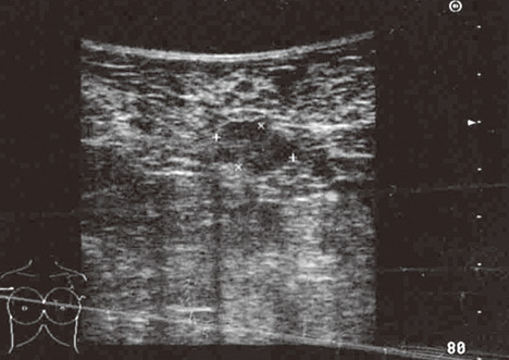
Figure 1 Ultrasonographic appearance of the breast mass. The mass is a hypoechogenic lesion with irregular margins measuring 4 cm in diameter.

Figure 2 Mammography of the right breast (mediolateral oblique view). Mammographic sensitivity decreased due to the increased density of the fibroglandular breast tissue.
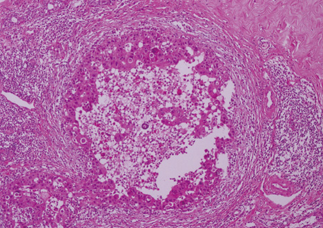
Figure 3 Histopathological appearance of high-grade ductal carcinoma in situ (H&E stain, × 200).
Reference
-
1. Amarasena R, Bowman S. Sjögren's syndrome. Clin Med. 2007. 7:53–56.
Article2. Kassan SS, Moutsopoulos HM. Clinical manifestations and early diagnosis of Sjögren syndrome. Arch Intern Med. 2004. 164:1275–1284.
Article3. Zhang W, Feng S, Yan S, Zhao Y, Li M, Sun J, et al. Incidence of malignancy in primary Sjögren's syndrome in a Chinese cohort. Rheumatology (Oxford). 2010. 49:571–577.
Article4. Lazarus MN, Robinson D, Mak V, Møller H, Isenberg DA. Incidence of cancer in a cohort of patients with primary Sjögren's syndrome. Rheumatology (Oxford). 2006. 45:1012–1015.
Article5. Yoshimura M, Koizumi K, Satani K, Kakizaki D, Kawanishi Y, Ohyashiki K, et al. Gallium-67 scintigraphic findings in a patient with breast lymphoma complicated with Sjögren syndrome. Ann Nucl Med. 2000. 14:227–229.
Article6. Boetes C, Mus RD, Holland R, Barentsz JO, Strijk SP, Wobbes T, et al. Breast tumors: comparative accuracy of MR imaging relative to mammography and US for demonstrating extent. Radiology. 1995. 197:743–747.
Article7. Ernster VL, Ballard-Barbash R, Barlow WE, Zheng Y, Weaver DL, Cutter G, et al. Detection of ductal carcinoma in situ in women undergoing screening mammography. J Natl Cancer Inst. 2002. 94:1546–1554.
Article8. Clinical practice guidelines in oncology - v.1: breast. National Comprehensive Cancer Network. 2009. Accessed March 8th, 2011. http://www.nccn.org/professionals/physician_gls/PDF/breast.pdf.
- Full Text Links
-
- Actions
-
Cited
- CITED
-
- Close
- Share
-
- Similar articles
-
- Ipsilateral Malignant Axillary Lymphadenopathy and Contralateral Reactive Lymph Nodes in a COVID-19 Vaccine Recipient With Breast Cancer
- Coronavirus disease 2019 (COVID-19) vaccination-induced unilateral axillary lymphadenopathy: case series with follow-up and literature review
- Management of Ipsilateral Axillary Lymphadenopathy Detected on Breast Ultrasound after COVID-19 Vaccination
- Efficacy of Ultrasound-Guided Core Needle Biopsy in Detecting Metastatic Axillary Lymph Nodes in Breast Cancer
- Axillary silicone lymphadenopathy caused by gel bleeding with intact silicone breast implants: a case report
