MRI Findings of Obstructed Hemivagina and Ipsilateral Renal Agenesis (OHVIRA syndrome) with a Blind Megaureter: Case Report
- Affiliations
-
- 1Department of Radiology, Korea University Anam Hospital, College of Medicine, Korea University, Seoul, Korea. urorad@korea.ac.kr
- KMID: 2175616
- DOI: http://doi.org/10.13104/imri.2015.19.3.196
Abstract
- Obstructed hemivagina and ipsilateral renal anomaly (OHVIRA) syndrome is an uncommon congenital abnormality of the female urogenital tract characterized by the triad of uterine didelphys, obstructed hemivagina, and ipsilateral renal agenesis. A 13-year-old female presented with acute lower abdominal pain. Magnetic resonance imaging (MRI) revealed uterine didelphys, hematometrocolpos, obstructed hemivagina, and right ipsilateral agenesis, consistent with OHVIRA syndrome. Also, a well-defined mass with fluid signal intensity, mimicking adnexal neoplasm was seen in the right lower pelvic cavity adjacent to the posterior wall of the bladder. Vaginal septotomy and drainage of hematometrocolpos were done initially, but unilateral hysterectomy was later performed to relieve the patient's symptoms. The cystic mass in the right lower pelvic cavity was also excised and confirmed as a blind megaureter.
MeSH Terms
Figure
-
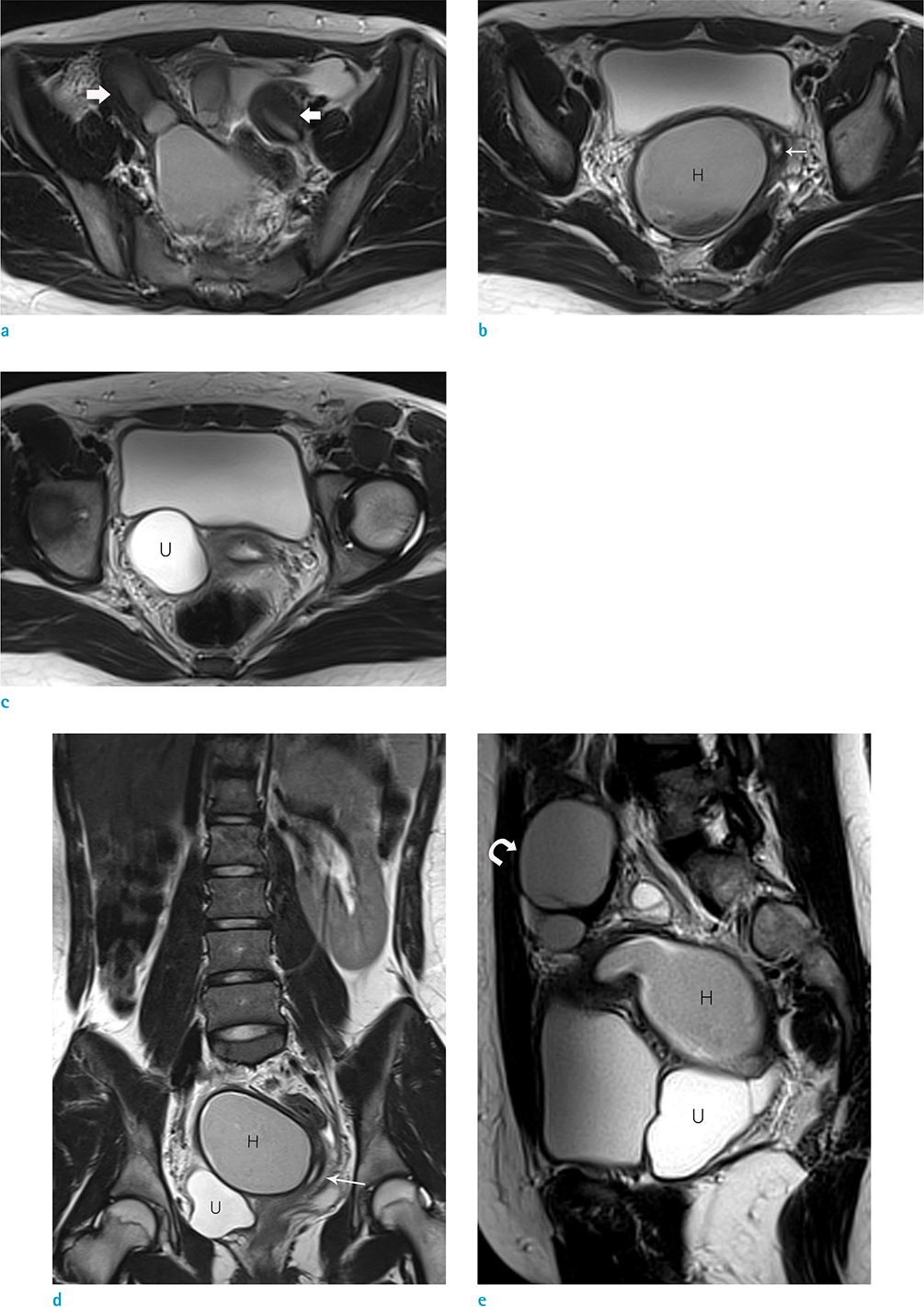
Fig. 1 Axial (a-c), coronal (d), and sagittal (e) turbo spin-echo T2-weighted images show two uterine horns (thick arrows), hematocolpos (H), hematosalpinx (curved arrow), and a blind megaureter (U). The right uterus was connected to the hematocolpos (H), and the left uterus and hemivagina (thin arrows) were seen on the left side of the hematocolpos (H). The right kidney was absent on the coronal turbo spin-echo T2-weighted image. A blind ectopic megaureter (U) was seen as a well-defined lobulated cystic mass posterior to the right aspect of the urinary bladder.
Cited by 1 articles
-
Two Cases of Herlyn-Werner-Wunderlich Syndrome in Neonates and Adolescents with Hydrocolpos and Hematocolpometra
Minjeong Park, Gyun-Ho Jeon
Perinatology. 2019;30(4):236-239. doi: 10.14734/PN.2019.30.4.236.
Reference
-
1. Lopes Dias J, Jogo R. Herlyn-Werner-Wunderlich syndrome: pre- and post-surgical MRI and US findings. Abdom Imaging. 2015; [Epub ahead of print].2. Orazi C, Lucchetti MC, Schingo PM, Marchetti P, Ferro F. Herlyn-Werner-Wunderlich syndrome: uterus didelphys, blind hemivagina and ipsilateral renal agenesis. Sonographic and MR findings in 11 cases. Pediatr Radiol. 2007; 37:657–665.3. Del Vescovo R, Battisti S, Di Paola V, et al. Herlyn-Werner-Wunderlich syndrome: MRI findings, radiological guide (two cases and literature review), and differential diagnosis. BMC Med Imaging. 2012; 12:4.4. Behr SC, Courtier JL, Qayyum A. Imaging of mullerian duct anomalies. Radiographics. 2012; 32:E233–E250.5. Smith NA, Laufer MR. Obstructed hemivagina and ipsilateral renal anomaly (OHVIRA) syndrome: management and follow-up. Fertil Steril. 2007; 87:918–922.6. Dobrocký I, Motoska V, Kemka S, Mistina lU. Ectopic blind ureter with ipsilateral renal agenesis: a case report. Eur J Radiol. 1995; 20:77–79.7. La Fianza A, Preda L, Di Maggio EM, Campani R. Blind megaureter with ipsilateral renal agenesis and mullerian anomaly: MR findings in a case. Clin Imaging. 1999; 23:184–186.8. Han B, Herndon CN, Rosen MP, Wang ZJ, Daldrup-Link H. Uterine didelphys associated with obstructed hemivagina and ipsilateral renal anomaly (OHVIRA) syndrome. Radiol Case Rep. 2010; 5:327.9. Donnez O, Jadoul P, Squifflet J, Donnez J. Didelphic uterus and obstructed hemivagina: recurrent hematometra in spite of appropriate classic surgical treatment. Gynecol Obstet Invest. 2007; 63:98–101.
- Full Text Links
-
- Actions
-
Cited
- CITED
-
- Close
- Share
-
- Similar articles
-
- Anatomical Variations, Genitourinary Anomalies and Clinical Presentations in Obstructed Hemivagina and Ipsilateral Renal Anomaly Syndrome: Case Series
- Magnetic resonance imaging in the evaluation of uterus didelphys with obstructed hemivagina and ipsilateral renal agenesis: a case report
- An unusual presentation of obstructed hemivagina and ipsilateral renal anomaly syndrome: A case report
- Uterus Didelphys with an Obstructed Hemivagina and Ipsilateral Renal Agenesis
- Complete septate uterus, obstructed hemivagina, and ipsilateral adnexal and renal agenesis in pregnancy
