Fast Cardiac CINE MRI by Iterative Truncation of Small Transformed Coefficients
- Affiliations
-
- 1Department of Electrical Engineering, Kwangwoon University, Seoul, Korea. cbahn@kw.ac.kr
- KMID: 2175573
- DOI: http://doi.org/10.13104/imri.2015.19.1.19
Abstract
- PURPOSE
A new compressed sensing technique by iterative truncation of small transformed coefficients (ITSC) is proposed for fast cardiac CINE MRI.
MATERIALS AND METHODS
The proposed reconstruction is composed of two processes: truncation of the small transformed coefficients in the r-f domain, and restoration of the measured data in the k-t domain. The two processes are sequentially applied iteratively until the reconstructed images converge, with the assumption that the cardiac CINE images are inherently sparse in the r-f domain. A novel sampling strategy to reduce the normalized mean square error of the reconstructed images is proposed.
RESULTS
The technique shows the least normalized mean square error among the four methods under comparison (zero filling, view sharing, k-t FOCUSS, and ITSC). Application of ITSC for multi-slice cardiac CINE imaging was tested with the number of slices of 2 to 8 in a single breath-hold, to demonstrate the clinical usefulness of the technique.
CONCLUSIONS
Reconstructed images with the compression factors of 3-4 appear very close to the images without compression. Furthermore the proposed algorithm is computationally efficient and is stable without using matrix inversion during the reconstruction.
Keyword
MeSH Terms
Figure
-

Fig. 1 Test data sets for cardiac CINE MRI for evaluation of the compressed sensing technique with other imaging methods.

Fig. 2 Three sampling strategies and corresponding sampling locations are exemplary shown for CF of 8: (a) uniform sampling, (b) Gaussian sampling, and (c) modified Gaussian sampling. The acquired locations are shown with white line segments at left, where the horizontal axis denotes the cardiac frame (1~16), and the vertical axis denotes the phase encoding gradient (-128~127). The corresponding histogram functions are shown at right, where the horizontal axis denotes the number of acquisitions over a period of the cardiac cycle, and the c vertical axis denotes the phase encoding gradient.
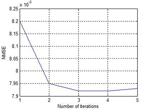
Fig. 3 NMSE of the reconstructed images by ITSC as a function of the number of iterations.
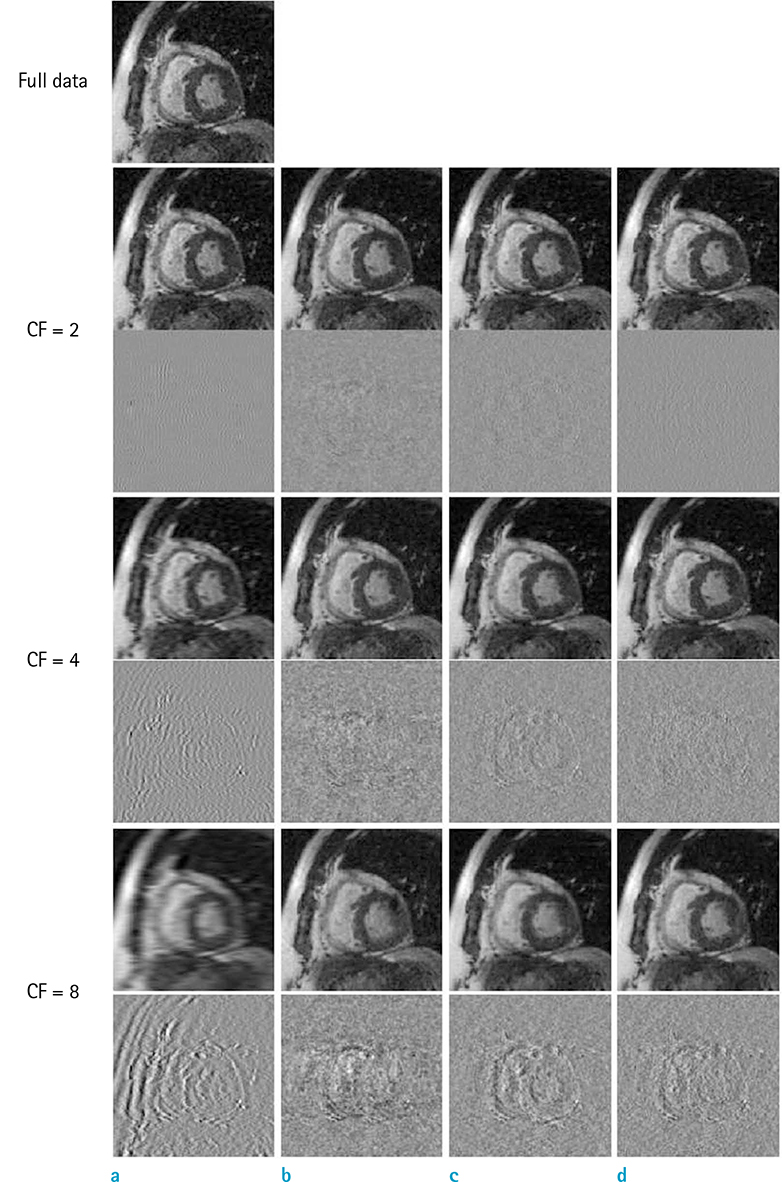
Fig. 4 Reconstructed images at a systolic phase, from the test data set A with CF of 2, 4, and 8 are shown for (a) zero filling, (b) view sharing, (c) k-t FOCUSS, and (d) ITSC. The conventionally reconstructed image with full data is shown at top. Error images are also shown for better visualization.
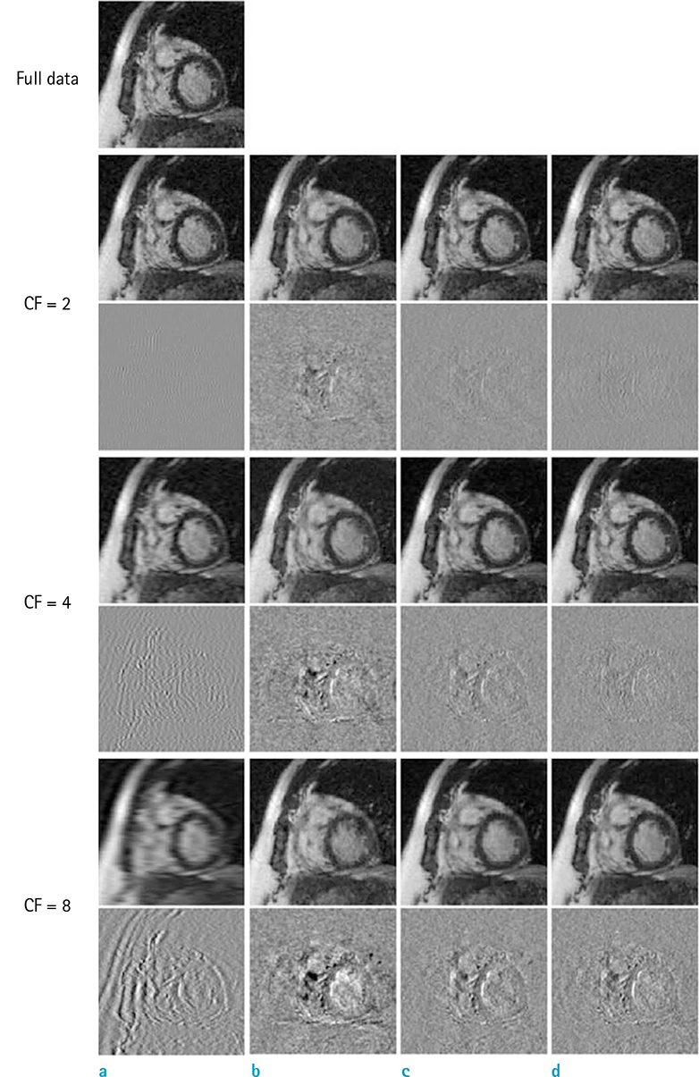
Fig. 5 Reconstructed images at a diastolic phase are shown for (a) zero filling, (b) view sharing, (c) k-t FOCUSS, and (d) ITSC with error images.
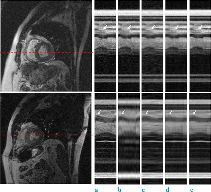
Fig. 6 The temporal profiles of the reconstructed images are shown right, for the test set A with CF of 4 (top) and for the test set B with CF of 8 (bottom). The horizontal axis is time, and the vertical axis is the broken line shown in the reconstructed images left corresponding to the phase encoding gradient direction for (a) reference with full data, (b) zero filling, (c) view sharing, (d) k-t FOCUSS, and (e) ITSC.
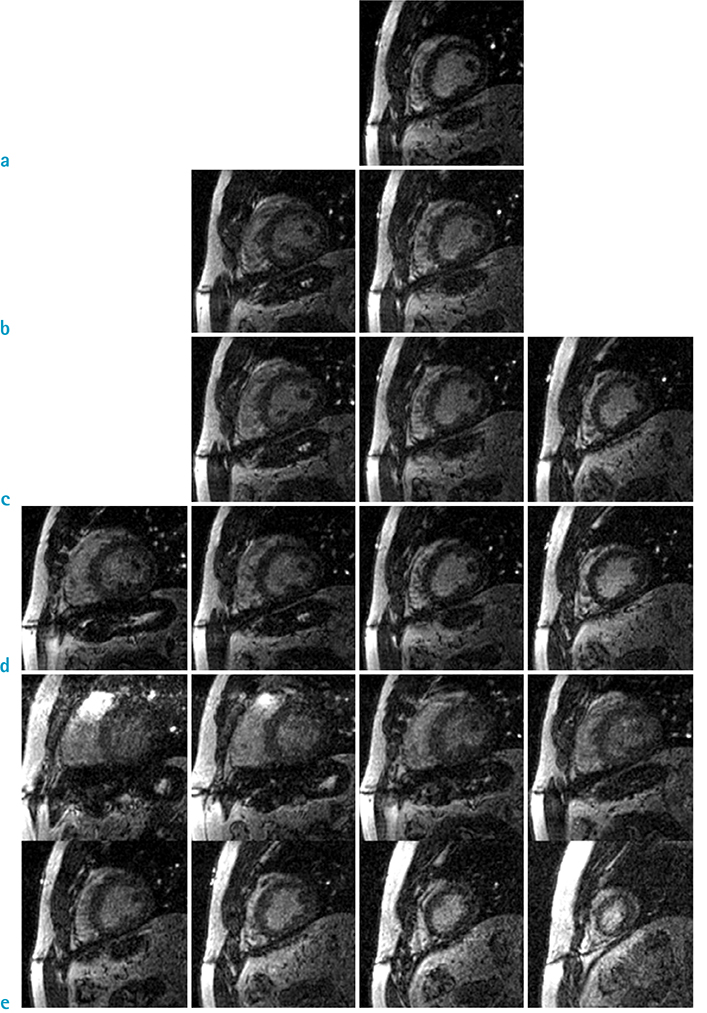
Fig. 7 In-vivo applications of ITSC for multi-slice cardiac CINE MRI. The reconstructed images are shown for (a) without compression, and with CF of (b) 2, (c) 3, (d) 4, and (e) 8. Note the number of slices obtained in single breath-hold is identical to the compression factor.
Cited by 1 articles
-
Biases in the Assessment of Left Ventricular Function by Compressed Sensing Cardiovascular Cine MRI
Jong-Hyun Yoon, Pan-ki Kim, Young-Joong Yang, Jinho Park, Byoung Wook Choi, Chang-Beom Ahn
Investig Magn Reson Imaging. 2019;23(2):114-124. doi: 10.13104/imri.2019.23.2.114.
Reference
-
1. Lee VS. Cardiovascular MRI: Physical Principles to Practical Protocols. Lippincott Williams &. Philadelphia: Lippincott Williams & Wilkins;2006.2. Carpenter TA, Williams EJ. MRI - from basic knowledge to advanced strategies: hardware. Eur Radiol. 1999; 9:1015–1019.3. Tsao J, Boesiger P, Pruessmann KP. k-t BLAST and k-t SENSE: dynamic MRI with high frame rate exploiting spatiotemporal correlations. Magn Reson Med. 2003; 50:1031–1042.4. Schmitt M, Potthast A, Sosnovik DE, et al. A 128-channel receive-only cardiac coil for highly accelerated cardiac MRI at 3 Tesla. Magn Reson Med. 2008; 59:1431–1439.5. Liu F, Zhao H, Crozier S. On the induced electric field gradients in the human body for magnetic stimulation by gradient coils in MRI. IEEE Trans Biomed Eng. 2003; 50:804–815.6. Glover PM. Interaction of MRI field gradients with the human body. Phys Med Biol. 2009; 54:R99–R115.7. Schoenberg SO, Dietrich O, Reiser MF. Parallel imaging in clinical MR applications. Berlin: Springer;2007.8. Riederer SJ, Tasciyan T, Farzaneh F, Lee JN, Wright RC, Herfkens RJ. MR fluoroscopy: technical feasibility. Magn Reson Med. 1988; 8:1–15.9. Doyle M, Walsh EG, Blackwell GG, Pohost GM. Block regional interpolation scheme for k-Space (BRISK): a rapid cardiac imaging technique. Magn Reson Med. 1995; 33:163–170.10. Jones RA, Haraldseth O, Müller TB, Rinck PA, Oksendal AN. K-space substitution: a novel dynamic imaging technique. Magn Reson Med. 1993; 29:830–834.11. Oesterle C, Strohschein R, Köhler M, Schnell M, Hennig J. Benefits and pitfalls of keyhole imaging, especially in first-pass perfusion studies. J Magn Reson Imaging. 2000; 11:312–323.12. Donoho DL. Compressed sensing. IEEE Trans Inf Theory. 2006; 52:1289–1306.13. Lustig M, Donoho D, Pauly JM. Sparse MRI: The application of compressed sensing for rapid MR imaging. Magn Reson Med. 2007; 58:1182–1195.14. Baraniuk RB. Compressive Sensing. IEEE Sign Proc Mag. 2007; 24:118–124.15. Jung H, Ye JC, Kim EY. Improved k-t BLAST and k-t SENSE using FOCUSS. Phys Med Biol. 2007; 52:3201–3226.16. Liang D, DiBella EV, Chen RR, Ying L. k-t ISD: dynamic cardiac MR imaging using compressed sensing with iterative support detection. Magn Reson Med. 2012; 68:41–53.17. Jung H, Ye JC. Motion estimated and compensated compressed sensing dynamic magnetic resonance imaging: What we can learn from video compression techniques. Int J Imaging Syst Technol. 2010; 20:81–98.18. Usman M, Atkinson D, Odille F, et al. Motion corrected compressed sensing for free-breathing dynamic cardiac MRI. Magn Reson Med. 2013; 70:504–516.19. Otazo R, Kim D, Axel L, Sodickson DK. Combination of compressed sensing and parallel imaging for highly accelerated first-pass cardiac perfusion MRI. Magn Reson Med. 2010; 64:767–776.20. Feng L, Grimm R, Block KT, et al. Golden-angle radial sparse parallel MRI: combination of compressed sensing, parallel imaging, and golden-angle radial sampling for fast and flexible dynamic volumetric MRI. Magn Reson Med. 2013; 72:707–717.21. Ahn CB. A New Compressed Sensing Technique by Iterative Truncation of Small Transformed Coefficients. Proc. ESMRMB. 2012. p. e-Poster 641.22. Bluemke DA, Boxerman JL, Atalar E, McVeigh ER. Segmented K-space cine breath-hold cardiovascular MR imaging: Part 1. Principles and technique. AJR Am J Roentgenol. 1997; 169:395–340.23. Park J, Yoon JH, Yang YJ, Ahn CB. Cardiac magnetic resonance imaging using Multi-physiological intelligent trigger system. J Korean Soc Magn Reson Med. 2014; 18:244–252.24. Margosian P. Faster MR imaging-imaging with half the data. Proc SMR. 1985; 1024–1025.25. Bernstein MA, King KF, Zhou XJ. Handbook of MRI Pulse Sequences. Amsterdam: Elsevier;2004.26. Lingala SG, Hu Y, DiBella E, Jacob M. Accelerated dynamic MRI exploiting sparsity and low-rank structure: k-t SLR. IEEE Trans Med Imaging. 2011; 30:1042–1054.27. Acharya T, Tsai PS. JPEG2000 Standard for Image Compression: Concepts, Algorithms and VLSI Architectures. New Jersey: John Wiley & Sons;2005.28. Gonzalez RC, Woods RE. Digital Image Processing. 3th ed. New Jersey: Pearson Education;2010.29. Vasanawala S, Murphy M, Alley M, et al. Practical parallel imaging compressed sensing MRI: Summary of two years of experience in acceleration body MRI of pediatric patients. Proc IEEE Int Symp Biomed Imaging. 2011; 1039–1043.30. Sharma SD, Fong CL, Tzung BS, Law M, Nayak KS. Clinical image quality assessment of accelerated magnetic resonance neuroimaging using compressed sensing. Invest Radiol. 2013; 48:638–645.
- Full Text Links
-
- Actions
-
Cited
- CITED
-
- Close
- Share
-
- Similar articles
-
- Fast Real-Time Cardiac MRI: a Review of Current Techniques and Future Directions
- Comparison between Echocardiography and Cardiac Cine-MRI : Left Ventricular Volume and Cardiac Output
- Comparison of Left and Right Ventricular Volume and Cardiac Output by MRI and Echocardiography
- Comparison of Cine Magnetic Resonance Imaging with Doppler Echocardiography for the Quantative Evaluation of Tricuspid Regurgitation in Newborn
- Cardiac MRI
