J Breast Cancer.
2009 Dec;12(4):338-343. 10.4048/jbc.2009.12.4.338.
Experience with Intracystic Papillary Carcinoma of the Breast at a Single Institute in Korea
- Affiliations
-
- 1Division of Breast and Endocrine Surgery, Department of Surgery, Samsung Medical Center, Sungkyunkwan University School of Medicine, Seoul, Korea. paojlus@hanmail.net
- 2Department of Pathology, Samsung Medical Center, Sungkyunkwan University School of Medicine, Seoul, Korea.
- KMID: 2175539
- DOI: http://doi.org/10.4048/jbc.2009.12.4.338
Abstract
- Intracystic papillary carcinoma (IPC) of the breast is a rare entity, which can be associated with ductal carcinoma in situ (DCIS) or an invasive carcinoma. There have been only limited reports describing IPC in Korea and the incidence among breast cancer cases in Korea has not yet been reported. From a database of 7,109 breast cancer cases treated surgically at Samsung Medical Center, we could only identify four IPC cases (0.056%). We also found that there are some differences between the clinicopathological characteristics of our cases and the alleged characteristics of IPC as reported from Western countries, such as a relatively young age of onset, small tumor size and various expression levels of hormonal receptors. We suspect this very low incidence may be caused by a true rarity of IPC in women in Korea or may be due to a lack of clinical interest for IPC. Upon presentation of our experience with IPC, we suggest the diagnosis for this rare disease entity needs to be reappraised.
MeSH Terms
Figure
-
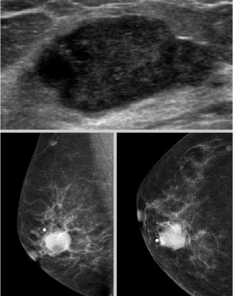
Figure 1 Mammography and Ultrasonography demonstrates a dense tumor with smooth and Iobulated contours which is the typical radiologic findings of intracystic papillary carcinoma.
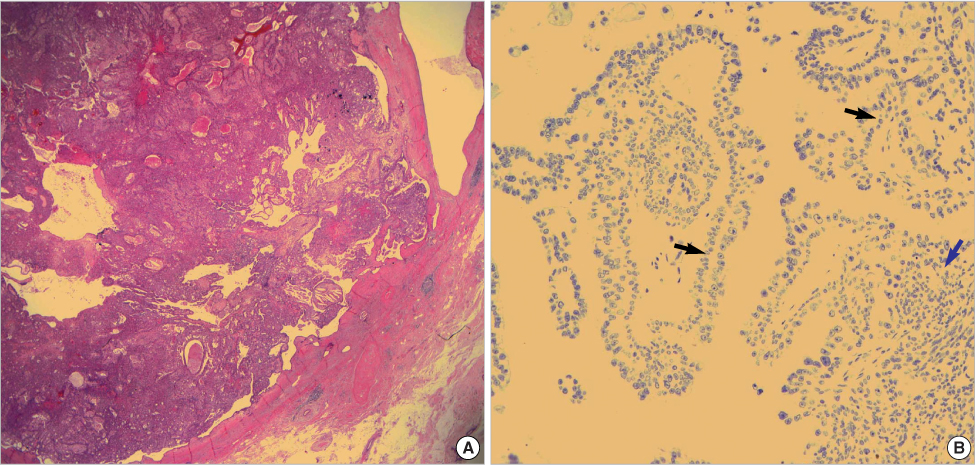
Figure 2 (A) Photomicrograph of tumor reveals an intracystic papillary carcinoma with thick fibrous capsule (H&E, ×10). (B) Immnohistochemistry for myoepithelial cells (p63) is negative at the periphery as well as at the cores of the papillae (×200) (black arrow: the core of the papillae, blue arrow: the periphery of the tumor).
Reference
-
1. Carter D, Orr SL, Merino MJ. Intracystic papillary carcinoma of the breast. After mastectomy, radiotherapy or excisional biopsy alone. Cancer. 1983. 52:14–19.
Article2. Leal C, Costa I, Fonseca D, Lopes P, Bento MJ, Lopes C. Intracystic (encysted) papillary carcinoma of the breast: a clinical, pathological, and immunohistochemical study. Hum Pathol. 1998. 29:1097–1104.
Article3. Lefkowitz M, Lefkowitz W, Wargotz ES. Intraductal (intracystic) papillary carcinoma of the breast and its variants: a clinicopathological study of 77 cases. Hum Pathol. 1994. 25:802–809.
Article4. Collins LC, Carlo VP, Hwang H, Barry TS, Gown AM, Schnitt SJ. Intracystic papillary carcinomas of the breast: a reevaluation using a panel of myoepithelial cell markers. Am J Surg Pathol. 2006. 30:1002–1007.
Article5. Yun SS, Choi SH, Kim SK, Park JK, Baek JM, Lee DH, et al. Intracystic papillary carcinoma in the male breast: a case report. J Breast Cancer. 2008. 11:151–155.
Article6. Woo OH, Yong HS, Kim A, Lee JB, Koo BH, Kang EY. Intracystic papillary carcinoma with extensive hemorrhage of the breast: sonographic and advanced MR findings: a case report. J Korean Radiol Soc. 2006. 55:511–514.
Article7. Park SC, Oh SJ, Kim KM, Kim JS, Jung SS. Case of an intracystic (encysted) papillary carcinoma of the breast. J Korean Surg Soc. 2000. 59:810–814.8. Ko KH, Kim EK, Park BW. Invasive papillary carcinoma of the breast presenting as post-traumatic recurrent hemorrhagic cysts. Yonsei Med J. 2006. 47:575–577.
Article9. Lee AW, Choi YJ, Lee KY, Kim BK, Kim SM, Shim SI. Fine needle aspiration cytology of intracystic papillary carcinoma of the breast. Korean J Cytopathol. 1997. 8:179–184.10. Collins LC, Schnitt SJ. Papillary lesions of the breast: selected diagnostic and management issues. Histopathology. 2008. 52:20–29.
Article11. Kraus FT, Neubecker RD. The differential diagnosis of papillary tumors of the breast. Cancer. 1962. 15:444–455.
Article12. Fisher ER, Palekar AS, Redmond C, Barton B, Fisher B. Pathologic findings from the National Surgical Adjuvant Breast Project (protocol no. 4). VI. Invasive papillary cancer. Am J Clin Pathol. 1980. 73:313–322.
Article13. Fayanju OM, Ritter J, Gillanders WE, Eberlein TJ, Dietz JR, Aft R, et al. Therapeutic management of intracystic papillary carcinoma of the breast: the roles of radiation and endocrine therapy. Am J Surg. 2007. 194:497–500.
Article14. Gendler LS, Feldman SM, Balassanian R, Riker MA, Frencher SK, Whelan DB, et al. Association of breast cancer with papillary lesions identified at percutaneous image-guided breast biopsy. Am J Surg. 2004. 188:365–370.
Article15. Viale G. Pathological definitions of invasion, metastatic potential and responsiveness to targeted therapies. Breast. 2007. 16:Suppl 2. S55–S58.
Article16. Mulligan AM, O'Malley FP. Metastatic potential of encapsulated (intracystic) papillary carcinoma of the breast: a report of 2 cases with axillary lymph node micrometastases. Int J Surg Pathol. 2007. 15:143–147.
Article17. Hill CB, Yeh IT. Myoepithelial cell staining patterns of papillary breast lesions: from intraductal papillomas to invasive papillary carcinomas. Am J Clin Pathol. 2005. 123:36–44.
Article18. Amemiya T, Oda K, Satake H, Ichihara S, Sawaki A, Shimoyama Y, et al. A case of intracystic papillary carcinoma accompanying widespread ductal carcinoma in situ. Breast Cancer. 2007. 14:312–316.
Article19. Solorzano CC, Middleton LP, Hunt KK, Mirza N, Meric F, Kuerer HM, et al. Treatment and outcome of patients with intracystic papillary carcinoma of the breast. Am J Surg. 2002. 184:364–368.
Article
- Full Text Links
-
- Actions
-
Cited
- CITED
-
- Close
- Share
-
- Similar articles
-
- Intracystic Papillary Carcinoma in the Male Breast: A Case Report
- Fine Needle Aspiration Cytology of Intracystic Papillary Carcinoma of the Breast
- Intracystic Papillary Carcinoma with Extensive Hemorrhage of the Breast: Sonographic and Advanced MR Findings: A Case Report
- Case of an Intracystic (Encysted) Papillary Carcinoma of the Breast
- Robotic Excision of a Huge Seminal Vesicle Cyst, Including Intracystic Papillary Adenoma, Saving Fertility
