The efficacy of sonographic morphology indexing and serum CA-125 for preoperative differentiation of malignant from benign ovarian tumors in patients after operation with ovarian tumors
- Affiliations
-
- 1Department of Obstetrics and Gynecology, College of Medicine, Chosun University, Gwangju, Korea. sjchoi@chosun.ac.kr
- KMID: 2173411
- DOI: http://doi.org/10.3802/jgo.2008.19.4.229
Abstract
OBJECTIVE
To evaluate the value of sonographic morphology indexing (MI) system and serum CA-125 levels in the assessment of the malignancy risk in patients with ovarian tumors.
METHODS
From September 2000 to July 2006, 202 patients who underwent surgery for ovarian tumors were reviewed retrospectively. In all patients, the MI score and serum CA-125 level were measured preoperatively. The association of the final pathologic diagnosis with the MI score and serum CA-125 level were examined.
RESULTS
There were 26 malignant tumors out of 141 ovarian tumors with a MI > or =5 (18%). With a cut-off value of 5, the sensitivity, specificity, PPV, and NPV of MI scores were 0.743, 0.293, 0.181, and 0.845, respectively. There were 22 malignant tumors out of 54 ovarian tumors with serum CA-125 >30 u/ml (41%). With a cut-off value of 30 u/ml, the sensitivity, specificity, PPV, and NPV of serum CA-125 level were 0.667, 0.808, 0.407, and NPV 0.925, respectively. On ROC curve, the optimal cut-off value of MI score was 6.5-7.5 and that of serum CA-125 level was 25.6-28.5 u/ml. With a cut-off value of 7, the sensitivity and 1-specificity of MI score were 0.875-0.917 and 0.023-0.203, respectively. After the exclusion of teratoma cases, the sensitivity and 1-specificity of MI score were 0.875-0.917 and 0.046-0.138, respectively. With a cut-off value of 25.6-28.5 u/ml, the sensitivity and 1-specificity of serum CA-125 level were 0.958 and 0.203-0.215, respectively.
CONCLUSION
The sonographic MI system is an accurate and simple method to differentiate a malignant tumor from a benign ovarian tumor. The accuracy of the sonographic MI system improved when the serum CA-125 level was considered and ovarian teratomas were excluded.
MeSH Terms
Figure
-
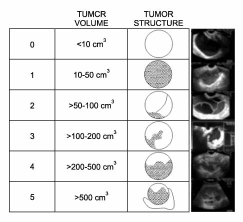
Fig. 1 Morphology indexing score.

Fig. 2 The ROC curve including ovarian teratomas. The cutoff range is a MI 6.5-7.5. It has a sensitivity 0.875-0.917 and 1-specificity 0.023-0.203. The cutoff range is CA-125 25.60-28.50. It has a sensitivity 0.958 and 1-specificity 0.203-0.215. MI: morphology indexing.
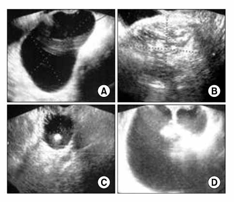
Fig. 3 Four sonographic findings of ovarian teratomas. Four types are a cystic pattern (A), a dense echo pattern with or without a cystic component (B), a pattern including a dense echogenic component with or without a cystic component (C), a densely echogenic tubercle associated with a cystic echo pattern (D).
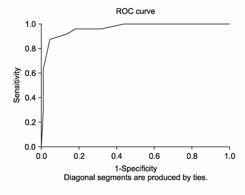
Fig. 4 The ROC curve excluding ovarian teratomas. A cutoff range is a MI 6.5-7.5. It has a sensitivity 0.875-0.917 and a 1-specificity 0.046-0.138. MI: morphology indexing.
Reference
-
1. Ministry of Health and Welfare Republic of Korea, Korea Central Cancer Registry. 2002 Annual report of the Korea central cancer registry (2002.1.1-2002.12.31). 2003. Seoul: Ministry of Health and Welfare Republic of Korea.2. Katsube Y, Berg JW, Silverberg SG. Epidemiologic pathology of ovarian tumors: A histopathologic review of primary ovarian neoplasms diagnosed in the Denver Standard Metropolitan Statistical Area, 1 July-31 December 1969 and 1 July-31 December 1979. Int J Gynecol Pathol. 1982. 1:3–16.3. Korean Society of Obstetrics and Gynecology. Annual report of gynecologic cancer registry program in Korea (2003.1.1-2003. 12.31). 2006. Seoul: Jin press.4. van Nagell JR Jr, DePriest PD, Reedy MB, Gallion HH, Ueland FR, Pavlik EJ, et al. The efficacy of transvaginal sonographic screening in asymptomatic women at risk for ovarian cancer. Gynecol Oncol. 2000. 77:350–356.5. Togashi K. MR imaging of the ovaries: normal appearance and benign disease. Radiol Clin North Am. 2003. 41:799–811.6. Scully RE, Young RH, Clement RB. Tumors of the ovary, maldeveloped gonads, fallopian tube, & broad ligament - 1998. 1998. Washington, DC: Armed Forces Institute of Pathology.7. Jacobs I, Oram D, Fairbanks J, Turner J, Frost C, Grudzinskas JG. A risk of malignancy index incorporating CA 125, ultrasound and menopausal status for the accurate preoperative diagnosis of ovarian cancer. Br J Obstet Gynaecol. 1990. 97:922–929.8. DePriest PD, Shenson D, Fried A, Hunter JE, Andrews SJ, Gallion HH, et al. A morphology index based on sonographic findings in ovarian cancer. Gynecol Oncol. 1993. 51:7–11.9. Pavlik EJ, DePriest PD, Gallion HH, Ueland FR, Reedy MB, Kryscio RJ, et al. Ovarian volume related to age. Gynecol Oncol. 2000. 77:410–412.10. Ueland FR, DePriest PD, Pavlik EJ, Kryscio RJ, van Nagell JR Jr. Preoperative differentiation of malignant from benign ovarian tumors: The efficacy of morphology indexing and Doppler flow sonography. Gynecol Oncol. 2003. 91:46–50.11. Mais V, Guerriero S, Ajossa S, Angiolucci M, Paoletti AM, Melis GB. Transvaginal ultrasonography in the diagnosis of cystic teratoma. Obstet Gynecol. 1995. 85:48–52.12. Partridge EE, Barnes MN. Epithelial ovarian cancer: Prevention, diagnosis, and treatment. CA Cancer J Clin. 1999. 49:297–320.13. DePriest PD, Varner E, Powell J, Fried A, Puls L, Higgins R, et al. The efficacy of a sonographic morphology index in identifying ovarian cancer: A multi-institutional investigation. Gynecol Oncol. 1994. 55:174–178.14. Sassone AM, Timor-Tritsch IE, Artner A, Westhoff C, Warren WB. Transvaginal sonographic characterization of ovarian disease: Evaluation of a new scoring system to predict ovarian malignancy. Obstet Gynecol. 1991. 78:70–76.15. Higgins RV, van Nagell JR Jr, Woods CH, Thompson EA, Kryscio RJ. Interobserver variation in ovarian measurements using transvaginal sonography. Gynecol Oncol. 1990. 39:69–71.16. Bailey CL, Ueland FR, Land GL, Depriest PD, Gallion HH, Kryscio RJ, et al. The malignant potential of small cystic ovarian tumors in women over 50 years of age. Gynecol Oncol. 1998. 69:3–7.17. Outwater EK, Siegelman ES, Hunt JL. Ovarian teratomas: Tumor types and imaging characteristics. Radiographics. 2001. 21:475–490.18. Quinn SF, Erickson S, Black WC. Cystic ovarian teratomas: The sonographic appearance of the dermoid plug. Radiology. 1985. 155:477–478.19. Choi JH, Tong SY, Kim MJ, Park JS, Lim YT, Kim JH, et al. Preoperative MR imaging for differentiation of immature from mature ovarian teratomas. Korean J Obstet Gynecol. 2006. 49:1547–1553.20. Grab D, Flock F, Stohr I, Nussle K, Rieber A, Fenchel S, et al. Classification of asymptomatic adnexal masses by ultrasound, magnetic resonance imaging, and positron emission tomography. Gynecol Oncol. 2000. 77:454–459.21. Kurtz AB, Tsimikas JV, Tempany CM, Hamper UM, Arger PH, Bree RL, et al. Diagnosis and staging of ovarian cancer: Comparative values of Doppler and conventional US, CT, and MR imaging correlated with surgery and histopathologic analysis--report of the Radiology Diagnostic Oncology Group. Radiology. 1999. 212:19–27.22. Royal College of Obstetricians and Gynaecologists. Ovarian cysts in postmenopausal women (Guideline; no. 34). 2003. London: Royal College of Obstetricians and Gynaecologists.23. Korean Society of Obstetrics and Gynecology. Gynecology. 2007. 4th ed. Seoul: Ko-Rye Press.24. Visintin I, Feng Z, Longton G, Ward DC, Alvero AB, Lai Y, et al. Diagnostic markers for early detection of ovarian cancer. Clin Cancer Res. 2008. 14:1065–1072.25. Mecke H, Savvas V. Laparoscopic surgery of dermoid cysts: Intraoperative spillage and complications. Eur J Obstet Gynecol Reprod Biol. 2001. 96:80–84.
- Full Text Links
-
- Actions
-
Cited
- CITED
-
- Close
- Share
-
- Similar articles
-
- Diagnostic Significance of Serum Tumor Markers in Paitents with Ovarian Tumors
- Significance of Serum CA 125 and CA 15-3 Levels in Patients with Ovarian Tumor
- A Study on Preoperative Diagnosis in Malignant Ovarian Tumor
- An Immunohistochemical Study of CA 125, CA 19-9, and CA 15-3 in Ovarian Epithelial Tumors
- Extremely elevated serum CA 125 in a borderline tumor of the ovary: A case report
