Ann Dermatol.
2011 Feb;23(1):111-114. 10.5021/ad.2011.23.1.111.
Branch-shaped Cutaneous Hypopigmentation and Atrophy after Intralesional Triamcinolone Injection
- Affiliations
-
- 1Department of Dermatology, College of Medicine, Chung-Ang University, Seoul, Korea. drseo@hanafos.com
- KMID: 2171968
- DOI: http://doi.org/10.5021/ad.2011.23.1.111
Abstract
- Cutaneous changes after local corticosteroid administration may include dermal atrophy, hyperpigmentation, alopecia, and hypopigmentation. Linear hypopigmentation and atrophy after intralesional injection of triamcinolone acetonide has been reported in the literature as a very rare side effect. A 30-year-old woman visited our dermatology department for a linear hypopigmented patch with atrophy from her left foot to the lower margin of the knee. The lesion developed after injection of an intralesional corticosteroid. The patient was diagnosed with linear hypopigmentation and atrophy secondary to the triamcinolone injection.
Keyword
MeSH Terms
Figure
-
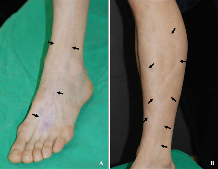
Fig. 1 (A) Hypopigmented patch with atrophy on the patient's left foot. (B) Linear hypopigmentation and atrophy along the left dorsal foot up to the lower margin of the knee.

Fig. 2 (A) The lesional side shows epidermal atrophy and flattening rete ridges (H&E, ×200). (B) The lesional side shows hypopigmentation of the basal layer (Fontana-Masson, ×200). (C) The normal side shows non-specific change (H&E, ×200). (D) In the normal side, pigmentation of the basal layer is not decreased (Fontana-Masson, ×200).
Reference
-
1. Firooz A, Tehranchi-Nia Z, Ahmed AR. Benefits and risks of intralesional corticosteroid injection in the treatment of dermatological diseases. Clin Exp Dermatol. 1995. 20:363–370.
Article2. Cantürk F, Cantürk T, Aydin F, Karagöz F, Sentürk N, Turanli AY. Cutaneous linear atrophy following intralesional corticosteroid injection in the treatment of tendonitis. Cutis. 2004. 73:197–198.3. Nanda V, Parwaz MA, Handa S. Linear hypopigmentation after triamcinolone injection: a rare complication of a common procedure. Aesthetic Plast Surg. 2006. 30:118–119.
Article4. George WM. Linear lymphatic hypopigmentation after intralesional corticosteroid injection: report of two cases. Cutis. 1999. 64:61–64.5. Schoepe S, Schäcke H, May E, Asadullah K. Glucocorticoid therapy-induced skin atrophy. Exp Dermatol. 2006. 15:406–420.
Article6. Gupta AK, Gover MD, Nouri K, Taylor S. The treatment of melasma: a review of clinical trials. J Am Acad Dermatol. 2006. 55:1048–1065.
Article7. Kikuchi I, Horikawa S. Perilymphatic atrophy of the skin. Arch Dermatol. 1975. 111:795–796.
Article8. Friedman SJ, Butler DF, Pittelkow MR. Perilesional linear atrophy and hypopigmentation after intralesional corticosteroid therapy. Report of two cases and review of the literature. J Am Acad Dermatol. 1988. 19:537–541.
Article9. Okere K, Jones MC. A case of skin hypopigmentation secondary to a corticosteroid injection. South Med J. 2006. 99:1393–1394.
Article
- Full Text Links
-
- Actions
-
Cited
- CITED
-
- Close
- Share
-
- Similar articles
-
- Intralesional Injection of Verapamil Only and Verapamil and Serial Triamcinolone Acetonide in Peyronie's Disease
- Giant Infantile Hemangioma Treated with Beta-blocker with Intermittent Triamcinolone Intralesional Injection
- Intralesional Injection of Triamcinolone for Urethral Stricture after Visual Urethrotomy
- Cutaneous Silicone Granuloma Treated with Oral Minocycline and Intralesional Injection of Triamcinolone Acetonide
- Hypertrophic Lupus Erythematosus Successfully Treated with Triamcinolone Intralesional Injection
