Scaly Ear Rash as the Herald of a Young Girl with Juvenile Systemic Lupus Erythematosus
- Affiliations
-
- 1Department of Dermatology, Tri-Service General Hospital, National Defense Medical Center, Taipei, Taiwan. jiangjienping@yahoo.com.tw
- KMID: 2171865
- DOI: http://doi.org/10.5021/ad.2011.23.S3.S333
Abstract
- Juvenile systemic lupus erythematosus (JSLE) is an autoimmune-mediated multiorgan disease. The cutaneous manifestation is one of the most common initial presentations in JSLE. A typical lesion is a facial malar rash, but a patient may sometimes present with nonclassical lesions. Herein, we report two cases of JSLE with similar persistent scaly ear rashes as the heralding cutaneous symptom preceding systemic symptoms. Identifying this atypical and underestimated cutaneous rash in juvenile patients might help the clinician make the correct diagnosis and provide earlier intervention, which may help prevent disease progression.
Keyword
Figure
-

Fig. 1 Patient 1. (A) Scaly erythematous plaque on the left ear. (B) Typical malar rah. (C) Painful ulcer on the hard palate. Patient 2. (D) Edematous, erythematous plaque with scales on the left ear. (E) Facial malar rash, discoid rash with petechiae. (F) Periungual erythema over the fingers.
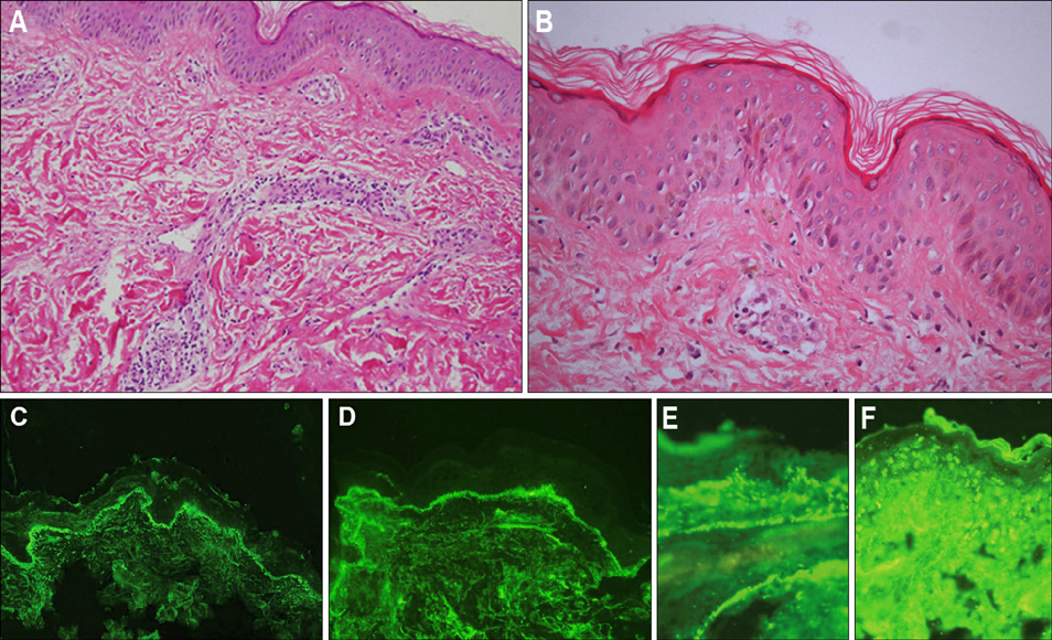
Fig. 2 Histological sections of skin biopsies, patient 1 (A) thickened basement membrane zone with dermal perivascular lymphocytic infiltrate (B) subtle vacuolar degeneration and melanin incontinence (H&E, original magnification, A: ×20, B: ×40), immunofluorescence staining revealing positive for lupus band (C) IgG and (D) IgM (original magnification, C: ×20, D: ×40). Patient 2 (E) positive lupus band of IgM and (F) nuclear staining of IgG (original magnification, D: ×20, F: ×20).
Reference
-
1. Hoffman IE, Lauwerys BR, De Keyser F, Huizinga TW, Isenberg D, Cebecauer L, et al. Juvenile-onset systemic lupus erythematosus: different clinical and serological pattern than adult-onset systemic lupus erythematosus. Ann Rheum Dis. 2009. 68:412–415.
Article2. Stichweh D, Arce E, Pascual V. Update on pediatric systemic lupus erythematosus. Curr Opin Rheumatol. 2004. 16:577–587.
Article3. Bader-Meunier B, Armengaud JB, Haddad E, Salomon R, Deschênes G, Koné-Paut I, et al. Initial presentation of childhood-onset systemic lupus erythematosus: a French multicenter study. J Pediatr. 2005. 146:648–653.
Article4. Gilliam JN, Sontheimer RD. Distinctive cutaneous subsets in the spectrum of lupus erythematosus. J Am Acad Dermatol. 1981. 4:471–475.
Article5. Werth VP. Clinical manifestations of cutaneous lupus erythematosus. Autoimmun Rev. 2005. 4:296–302.
Article6. Watanabe T, Tsuchida T. Classification of lupus erythematosus based upon cutaneous manifestations. Dermatological, systemic and laboratory findings in 191 patients. Dermatology. 1995. 190:277–283.
Article7. Albrecht J, Werth VP. Clinical outcome measures for cutaneous lupus erythematosus. Lupus. 2010. 19:1137–1143.
Article8. Vyas S, Hidalgo G, Baqi N, Von Gizyki H, Singh A. Outcome in African-American children of neuropsychiatric lupus and lupus nephritis. Pediatr Nephrol. 2002. 17:45–49.
Article9. Bernatsky S, Boivin JF, Joseph L, Manzi S, Ginzler E, Gladman DD, et al. Mortality in systemic lupus erythematosus. Arthritis Rheum. 2006. 54:2550–2557.
Article10. Marks SD, Tullus K. Modern therapeutic strategies for paediatric systemic lupus erythematosus and lupus nephritis. Acta Paediatr. 2010. 99:967–974.
Article
- Full Text Links
-
- Actions
-
Cited
- CITED
-
- Close
- Share
-
- Similar articles
-
- Multiple Eruptive Dermatofibromas in a Patient with Systemic Lupus Erythematosus and Juvenile Rheumatoid Arthritis
- Sequential Development of Systemic Lupus Erythematosus in a Patient with Juvenile Rheumatoid Arthritis
- A Case of Chilblain Lupus Erythematosus
- A Case Of Systemic Lupus Erythematosus Associated With Hyperthyroidism And Severe Retinopathy
- A Case of Chilblain Lupus Erythematosus
