Rapid Growing Superficial Cutaneous Leiomyosarcoma of the Face
- Affiliations
-
- 1Department of Dermatology, School of Medicine, Chosun University, Gwangju, Korea. derm75@chosun.ac.kr
- 2Department of Orthopedic Surgery, School of Medicine, Chosun University, Gwangju, Korea.
- KMID: 2171797
- DOI: http://doi.org/10.5021/ad.2013.25.2.237
Abstract
- Leiomyosarcomas are uncommon malignant smooth muscle tumors, mainly derived from vessels or viscera. Superficial leiomyosarcomas are a rare soft tissue sarcoma arising from the dermis or subcutaneous tissue in the skin. According to tumor origin and location, they are divided into cutaneous and subcutaneous leiomyosarcoma. They have distinctly different histologic and prognostic features from each other. Superficial leiomyosarcomas show a predilection for the proximal extremities and tend to be slow growing. We report one rare case of superficial cutaneous leiomyosarcoma on the right temporal area of face, which showed an extremely rapid growing mass within 3 months.
Keyword
Figure
-
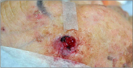
Fig. 1 Solitary 2×3 cm, well-demarcated, soft erythematous nodule with ulceration and bleeding on right temporal area of face.
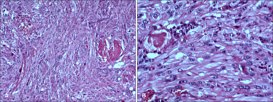
Fig. 2 Histopathologic findings of the lesion revealed interlacing fascicles of spindle-shaped cells with varying degrees of intermingled collagen. Spindle cells had elongated blunt-ended nuclei with mitosis and hyperchromatic nuclei (H&E stain; A: ×100, B: ×200).

Fig. 3 Results of immunohistochemical staining showed a positive reaction of actin (A), vimentin (B), and desmin (C), and a negative reaction of CD-34 (D), epithelial membrane antigen (EMA) (E), cytokeratin (F), factor VIII (G), and S-100 (H) (A~H: ×100).
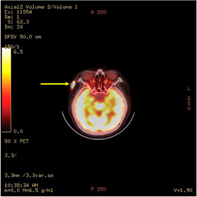
Fig. 4 Positron emission tomography-computed tomography showed a hypermetabolic lesion (arrow) on the right temporal area of the head.
Reference
-
1. Holst VA, Junkins-Hopkins JM, Elenitsas R. Cutaneous smooth muscle neoplasms: clinical features, histologic findings, and treatment options. J Am Acad Dermatol. 2002. 46:477–490.
Article2. Torres T, Oliveira A, Sanches M, Selores M. Superficial cutaneous leiomyosarcoma of the face: report of three cases. J Dermatol. 2011. 38:373–376.
Article3. De Giorgi V, Sestini S, Massi D, Papi F, Alfaioli B, Lotti T. Superficial cutaneous leiomyosarcoma: a rare, misleading tumor. Am J Clin Dermatol. 2008. 9:185–187.4. Lin JY, Tsai RY. Subcutaneous leiomyosarcoma on the face. Dermatol Surg. 1999. 25:489–491.
Article5. Blaise G, Nikkels AF, Quatresooz P, Hermanns-Lê T, Piérard GE. Childhood cutaneous leiomyosarcoma. Pediatr Dermatol. 2009. 26:477–479.
Article6. Choy C, Cooper A, Kossard S. Primary cutaneous diffuse leiomyosarcoma with desmoplasia. Australas J Dermatol. 2006. 47:291–295.
Article7. Elder DE, Elenitsas R, Johnson BL Jr, Murphy GF, Xu G. Lever's histopathology of the skin. 2009. 10th ed. Philadelphia: Wolters Kluwer/Lippincott Williams & Wilkins Cop..8. Annest NM, Grekin SJ, Stone MS, Messingham MJ. Cutaneous leiomyosarcoma: a tumor of the head and neck. Dermatol Surg. 2007. 33:628–633.
Article9. Tsutsumida A, Yoshida T, Yamamoto Y, Itoh T, Minakawa H, Sugihara T. Management of superficial leiomyosarcoma: a retrospective study of 10 cases. Plast Reconstr Surg. 2005. 116:8–12.
Article10. Fields JP, Helwig EB. Leiomyosarcoma of the skin and subcutaneous tissue. Cancer. 1981. 47:156–169.
Article11. Kim DY, Song JY. A case of primary cutaneous leiomyosarcoma. Korean J Dermatol. 1984. 22:445–448.12. Na GY, Sung GY, Jun JB, Suh SB. A case of primary cutaneous leiomyosarcoma. Korean J Dermatol. 1988. 26:106–109.13. Park HS, Kim SJ, Choi JC, Chun DK, Lee YS. A case of cutaneous leiomyosarcoma on the face. Korean J Dermatol. 2003. 41:1111–1113.14. Rho JH, Choi HB, Joh OJ, Park SR, Song KY. A case of cutaneous leiomyosarcoma. Korean J Dermatol. 2005. 43:121–123.15. Chun SH, Jeon SY, Lee WS. A case of cutaneous leiomyosarcoma on the sole. Korean J Dermatol. 2005. 43:707–709.16. Park KY, Han TY, Seo SJ, Hong CK, Song KY. A case of dermal type of cutaneous leiomyosarcoma. Korean J Dermatol. 2007. 45:938–941.17. Lee HE, Jue MS, Ko JY, Kim YH, Ro YS. A case of uterine leiomyosarcoma metastasized to the skin: local recurrence and rapid growth after excision. Korean J Dermatol. 2010. 48:346–349.
