A Case Report of Hajdu-Cheney Syndrome
- Affiliations
-
- 1Department of Endocrinology and Metabolism, Ajou University School of Medicine, Suwon, Korea. yschung@ajou.ac.kr
- 2Department of Radiology, Ajou University School of Medicine, Suwon, Korea.
- KMID: 2169023
- DOI: http://doi.org/10.3803/EnM.2010.25.2.152
Abstract
- Hajdu-Cheney syndrome (HCS) is a rare skeletal dysplasia that is characterized by acroosteolysis of the distal phalanges, distinctive craniofacial and skull changes, dental abnormalities and generalized osteoporosis. The clinical and radiologic characteristics are variable and these characteristics progress with age. This syndrome shows autosomal dominant inheritance with sporadic cases. The genetic defects or molecular pathogenesis of HCS are still unknown. We experienced a case of Hajdu-Cheney syndrome in a 20-year-old man who had generalized osteoporosis with multiple non-traumatic spine compression fractures. He had acroosteolysis of the hands and feet, wormian bones in the skull, facial dysmorphism (mid-facial flattening, micrognathia and bushy eyebrows), a high arched palate, malocclusion and short dental alveolar processes. HCS was diagnosed based on the clinical and radiologic evidence. For the differential diagnosis, we excluded the other possible causes of the acroosteolysis and wormian bones, including hyperparathyroidism, osteogenesis imperfecta, hypophosphatemia and mandibuloacral dysplasia. The specific treatment of HCS is unknown, but case reports with bisphosphonate treatment have been reported.
MeSH Terms
Figure
-

Fig. 1 Clinical photograph of face*. Coarse face and hair, bushy eyebrow and micrognathia are observed. *This facial photograph is submitted with informed consent of the patient for academic purpose.

Fig. 2 Clinical photo of hands. Short and clubbed fingers due to acroosteolysis.
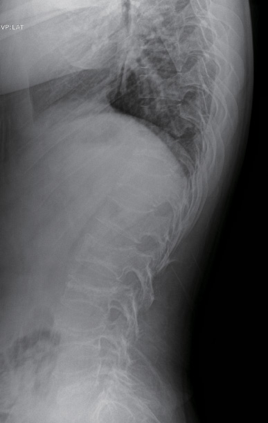
Fig. 3 Thoracolumbar spine lateral projection shows osteoporosis with biconcave vertebral bodies due to compression fractures.

Fig. 4 Phalangeal acroosteolysis of hands (A) and feet (B). The osteolysis is most marked in the middle of the distal phalanges, ranging from loss of part of the tuft to loss of almost entire distal phalanges. Note cone-shaped distal phalanges of the toes due to peripheral acrolyses.

Fig. 5 Persistence of coronal suture into adult life and wormian bones in the lambdoidal suture are seen.
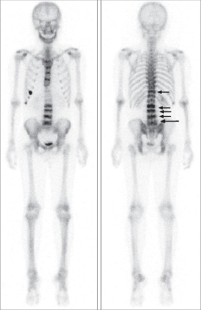
Fig. 6 Multiple compression fractures of spine. 99mTc-MDP whole body bone scan revealed multiple compression fractures in T11 and L2-L5 spines (arrows) and focal uptakes in right 6th, 7th and 8th costochondral junctions.
Cited by 2 articles
-
Effect of Zoledronic Acid on Acro-Osteolysis and Osteoporosis in a Patient with Hajdu-Cheney Syndrome
Sena Hwang, Dong Yoeb Shin, Seong Hwan Moon, Eun Jig Lee, Sung-Kil Lim, Ok Hwa Kim, Yumie Rhee
Yonsei Med J. 2011;52(3):543-546. doi: 10.3349/ymj.2011.52.3.543.Band acro-osteolysis in a Black woman: a case report and review of the literature
Jin-Myoung Dan, Cheungsoo Ha, Ho-Jae Lee
Arch Hand Microsurg. 2022;27(1):62-67. doi: 10.12790/ahm.21.0140.
Reference
-
1. Kim OH, Nishimura G. Congenital Skeletal Malformation syndromes. 2006. Seoul: Ryomoongak;97–99.2. Hajdu N, Kauntze R. Cranio-skeletal dysplasia. Br J Radiol. 1948. 21:42–48.3. Cheney WD. Acro-osteolysis. Am J Roentgenol Radium Ther Nucl Med. 1965. 94:595–607.4. Brennan AM, Pauli RM. Hajdu--Cheney syndrome: evolution of phenotype and clinical problems. Am J Med Genet. 2001. 100:292–310.5. Shin DY, Kim KM, Ku CR, Choi HS, Rhee YM, Moon SH, Park YK, Lee EJ, Kim OH, Lim SK. A Case of Hadju-Cheney Syndrome diagnosed as acroosteolysis of Hands and Foot. J Korean Soc osteoporos. 2007. 5:S5. 271.6. Marik I, Kuklik M, Zemkowa D, Kozlowski K. Hajdu-Cheney syndrome: report of a family and a short literature review. Australas Radiol. 2006. 50:534–538.7. Currarino G. Hajdu-Cheney syndrome associated with serpentine fibulae and polycystic kidney disease. Pediatr Radiol. 2009. 39:47–52.8. McKiernan FE. Integrated anti-remodeling and anabolic therapy for the osteoporosis of Hajdu-Cheney syndrome. Osteoporos Int. 2007. 18:245–249.9. O'Reilly M, Shaw DG. Hajdu-Cheney syndrome. Ann Rheum Dis. 1994. 53:276–279.10. Iwaya T, Taniguchi K, Watanabe J, Iinuma K, Hamazaki Y, Yoshikawa S. Hajdu-Cheney syndrome. Arch Orthop Trauma Surg. 1979. 95:293–302.11. Brown DM, Bradford DS, Gorlin RJ, Desnick RJ, Langer LO, Jowsey J, Sauk JJ. The acro-osteolysis syndrome: morphologic and biochemical studies. J Pediatr. 1976. 88:573–580.12. Elias AN, Pinals RS, Anderson HC, Gould LV, Streeten DH. Hereditary osteodysplasia with acro-osteolysis. (The Hajdu-Cheney syndrome). Am J Med. 1978. 65:627–636.13. Nunziata V, Di Giovanni G, Ballanti P, Bonucci E. High turnover osteoporosis in acro-osteolysis (Hajdu-Cheney syndrome). J Endocrinol Invest. 1990. 13:251–255.14. Leidig-Bruckner G, Pfeilschifter J, Penning N, Limberg B, Priemel M, Delling G, Ziegler R. Severe osteoporosis in familial Hajdu-Cheney syndrome: progression of acro-osteolysis and osteoporosis during long-term follow-up. J Bone Miner Res. 1999. 14:2036–2041.15. Kamath RP, Chandran P, Ankarath S. Musculoskeletal manifestations of Hajdu-Cheney syndrome. Eur J Radiol Extra. 2005. 54:83–86.16. Drake WM, Hiorns MP, Kendler DL. Hadju-Cheney syndrome: response to therapy with bisphosphonates in two patients. J Bone Miner Res. 2003. 18:131–133.17. Gökçe C, Gökçe O, Baydinç C, Ilhan N, Alaşehirli E, Ozküçük F, Taşçi M, Atilkeler MK, Celebi H, Arslan N. Use of random urine samples to estimate total urinary calcium and phosphate excretion. Arch Intern Med. 1991. 151:1587–1588.18. Nordin BE. Assessment of calcium excretion from the urinary calcium/creatinine ratio. Lancet. 1959. 274:368–371.
- Full Text Links
-
- Actions
-
Cited
- CITED
-
- Close
- Share
-
- Similar articles
-
- Effect of Zoledronic Acid on Acro-Osteolysis and Osteoporosis in a Patient with Hajdu-Cheney Syndrome
- An Unusual Presentation of Diabetic Ketoacidosis in Familial Hajdu-Cheney Syndrome: A Case Report
- The Eosinophilia-Myalgia Syndrome not Associated with L-tryptophan: A case report
- Thenar Compartment syndrome: A Case Report
- A Case of Chronic Fatigue Syndrome: Case Report & Literature Review
