Shape and innervation of popliteus muscle
- Affiliations
-
- 1Department of Plastic Surgery, and Center for Advanced Medical Education by BK21 Project, Inha University School of Medicine, Incheon, Korea. jokerhg@inha.ac.kr
- 2Department of Physical Medicine and Rehabilitation, Chungbuk University Medical School, Chungju, Korea.
- 3Department of Anatomy and Institute for Applied Anatomy, The Catholic Universty of Korea, Seoul, Korea.
- 4Department of Plastic Surgery, Gacheon Gil Hospital, Incheon, Korea.
- KMID: 2168890
- DOI: http://doi.org/10.5115/acb.2010.43.2.165
Abstract
- The aim of this study was to delineate the shape of the popliteus muscle and determine the correct motor point site for treating spasticity. A total of 22 legs from 13 fresh Korean cadavers were evaluated. The x-axis was set as a transverse line across the lateral and medial epicondyle of the femur and the y-axis as a vertical line at the midpoint of the medial malleolus of the tibia and lateral malleolus of the fibula. The popliteus muscle is an obtuse triangle in shape. Superior, medial, and inferior angles were 27.2+/-4.3degrees, 114.8+/-19.8degrees, and 38.0+/-18.8degrees respectively. The lengths of the superior, medial, and lateral sides of the triangle were 7.6+/-1.0 cm, 6.2+/-1.0 cm, and 11.9+/-1.5 cm respectively. Nerve branches ran superficially on the periosteum of the tibia and entered the popliteus on its superficial surface. The diverging point of the nerve branch entered the popliteus from the tibial nerve located at the midline of the popliteal fossa and 17% of the leg length above the intercondylar line. Most nerve entry points (83.3%) were within a 2.0x3.0 cm rectangle with the center located at -1.0 cm (-7%) on the x-axis and -3.3 cm (-9%) on the y-axis.
Keyword
Figure
-
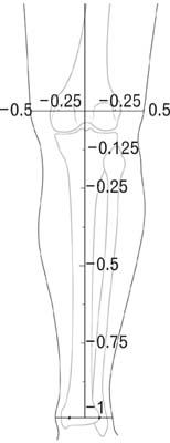
Fig. 1 Rectangular coordinates: x-axis crosses over the lateral and medial epicondyle of the femur; y-axis accords with a vertical mid-line between the medial malleous of the tibia and lateral malleolus of the fibula.
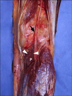
Fig. 2 Diverging point (arrow) of the nerve branch to the popliteus from the tibial nerve: nerve entry points (arrow heads) in the popliteus muscle (P).
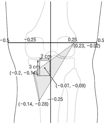
Fig. 3 Triangular popliteus: superior, medial, and inferior angles were 27.2±4.3°, 114.8±19.8°, and 38.0±18.8° respectively; superior, medial, and lateral sides of the triangle were 7.6±1.0 cm, 6.2±1.0 cm, and 11.9 ±1.5 cm respectively.

Fig. 4 Most nerve entry points (83.3%) were within a 2.0 by 3.0 cm rectangle with the center located at -1.0 cm (-7%) on the x-axis and -3.3 cm (-9%) on the y-axis.
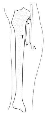
Fig. 5 Nerve branches (arrow heads) running superficially on the periosteum of the tibia entering the popliteus on its superficial surface about 1 cm distal to its superior border. T: tibia, P: popliteus, TN: tibial nerve.
Reference
-
1. Glenn MB. Glenn MB, Whyte J, editors. Nerve blocks. The practical management of spasticity in children and adults. 1990. Philadelphia: Lea & Febiger;227–258.2. Hollinshed WH. Functional anatomy of the limbs and back. 1986. 4th ed. Philadelphia: W.B. Saunders Co.;318–322.3. Kong KH, Chua KS. Intramuscular neurolysis with alcohol to treat post-stroke finger flexor spasticity. Clin Rehabilit. 2002. 16:378–381.4. Lehmann JF, De Lateur BJ. Kottke FJ, Lehmann JF, editors. Gait analysis: diagnosis and management. Krusen's handbook of physical medicine and rehabilitation. 1990. 4th ed. Philadelphia: W.B. Saunders Co.;108–111.5. Li YH, Leong JC. Intoeing gait in children. Hong Kong Med J. 1999. 5:360–366.6. Mann RA, Hagy JL. The popliteus muscle. J Bone Joint Surg Am. 1977. 59:924–927.7. Rethlefsen SA, Healy BS, Wren TA, Skaggs DL, Kay RM. Causes of intoeing gait in children with cerebral palsy. J Bone Joint Surg Am. 2006. 88:2175–2180.8. Sass P, Hassan G. Lower extremity abnormalities in children. Am Fam Physician. 2003. 68:461–468.9. Standring S. Gray's anatomy. 2005. 39th ed. Edinburgh: Elsevier;1485.
- Full Text Links
-
- Actions
-
Cited
- CITED
-
- Close
- Share
-
- Similar articles
-
- MRI Findings of Accessory Popliteus Muscle: A Case Report
- Extensor Digitorum Brevis Innervated by the Tibial Nerve (All Tibial Foot): A case report
- Neurovascular Compression Caused by Popliteus Muscle Enlargement Without Discrete Trauma
- Nerve-sparing radical hysterectomy: time for a new standard of care for cervical cancer?
- Diagnostic Values of SPACE Test in Corpus Cavernous Smooth Muscle
