Florid osseous dysplasia in a middle-aged Turkish woman: A case report
- Affiliations
-
- 1Department of Dentomaxillofacial Radiology, Faculty of Dentistry, Ankara University, Ankara, Turkey. dtbuketonder@gmail.com
- 2Department of Oral Pathology, Faculty of Dentistry, Gazi University, Ankara, Turkey.
- 3Department of Dentomaxillofacial Surgery, Faculty of Dentistry, Ankara University, Ankara, Turkey.
- KMID: 2167458
- DOI: http://doi.org/10.5624/isd.2013.43.3.197
Abstract
- Florid osseous dysplasia (FOD) is an uncommon, benign, cemento-osseous lesion of the jaws. The etiology of FOD is still unknown. It is often asymptomatic and may be identified on routine dental radiographs. The classic radiographic appearance of FOD is amorphous, lobulated, mixed radiolucent/radiopaque masses of cotton-wool appearance with a sclerotic border in the jaws. In our case the lesion was found incidentally on routine periapical radiographs taken for restored teeth and edentulous areas. For further and detailed examination, a panoramic radiograph and cone-beam computed tomograph (CBCT) were taken. The panoramic radiograph and CBCT revealed maxillary bilateral and symmetrical, non-expansile, well-defined, round, radiopaque masses in contact with the root of the maxillary right second molar and left first molar teeth. Our aim in presenting this case report was to highlight the importance of imaging in diagnosis of FOD.
Keyword
Figure
-
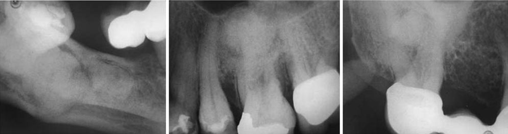
Fig. 1 Periapical radiographs reveal dense, amorphous, radiopaque masses.
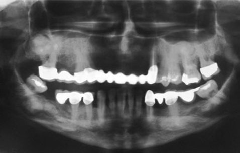
Fig. 2 A panoramic radiograph shows the mixed radiolucent/radiopaque masses with a cotton-wool appearance and with the entire border in three quadrants.
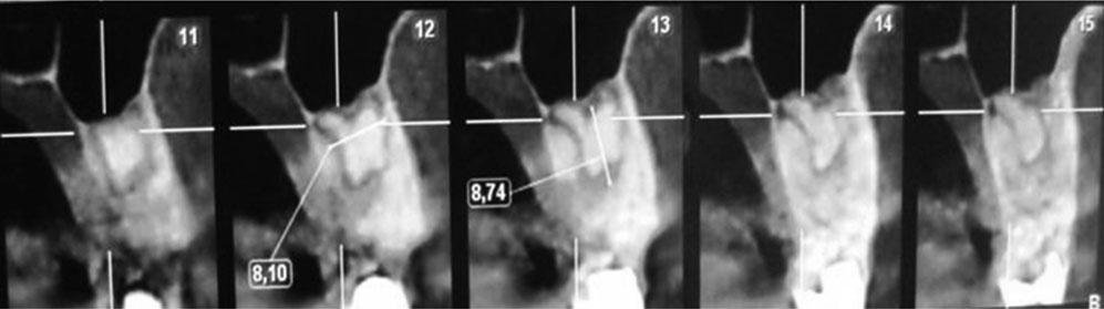
Fig. 3 CBCT cross-sectional images reveal the relationship between the lesion and the structures around the lesion, which is a radiopaque conglomerate separate from the bone with a radiolucent border.

Fig. 4 An axial CBCT image shows high-density, lobulated, well-defined, expansile masses of florid osseous dysplasias in the maxilla (arrows).
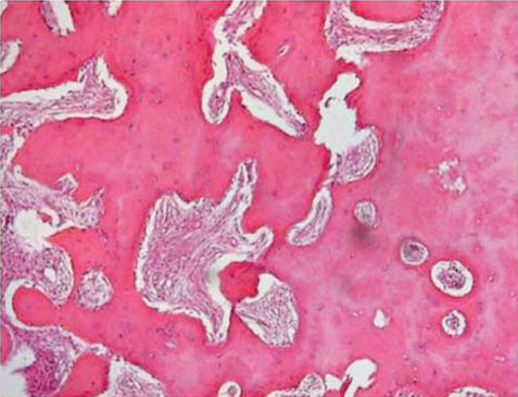
Fig. 5 Histopathological examination shows calcified cells within a fibrous matrix (H&E stain, 40×).
Cited by 1 articles
-
Recurrent symptomatic cemento-osseous dysplasia: A case report
Chang-Ki Min, Kwang-Joon Koh, Kyoung-A Kim
Imaging Sci Dent. 2018;48(2):131-137. doi: 10.5624/isd.2018.48.2.131.
Reference
-
1. White SC, Pharoah MJ. Oral radiology: principles and interpretation. 6th ed. St. Louis: Mosby/Elsevier;2000. p. 437.2. Ong ST, Siar CH. Florid cemento-osseous dysplasia in a young Chinese man. Case report. Aust Dent J. 1997; 42:404–408.
Article3. Barnes L, Eveson JW, Reichart P, Sidransky D. Pathology and genetics of head and neck tumours. World Health Organization Classification of tumours. Lyon: IARC Press;2005. p. 323.4. Gonçalves M, Píspico R, Alves Fde A, Lugão CE, Gonçalves A. Clinical, radiographic, biochemical and histological findings of florid cemento-osseous dysplasia and report of a case. Braz Dent J. 2005; 16:247–250.
Article5. Dağıstan S, Tozoğlu Ü, Göregen M, Çakur B. Florid cemento-osseous dysplasia: a case report. Med Oral Patol Oral Cir Bucal. 2007; 12:E348–E350.6. MacDonald-Jankowski DS. Florid cemento-osseous dysplasia: a systematic review. Dentomaxillofac Radiol. 2003; 32:141–149.
Article7. Gündüz K, Avsever H, Karaçaylı Ü, Senel B, Pişkin B. Florid cemento-osseous dysplasia: a case report. Braz Dent J. 2009; 20:347–350.
Article8. Singer SR, Mupparapu M, Rinaggio J. Florid cemento-osseous dysplasia and chronic diffuse osteomyelitis: report of a simultaneous presentation and review of the literature. J Am Dent Assoc. 2005; 136:927–931.9. Minhas G, Hodge T, Gill DS. Orthodontic treatment and cemento-osseous dysplasia: a case report. J Orthod. 2008; 35:90–95.
Article10. Jerjes W, Banu B, Swinson B, Hopper C. Florid cemento-osseous dysplasia in a young Indian woman. A case report. Br Dent J. 2005; 198:477–478.
Article11. Worawongvasu R, Songkampol K. Fibro-osseous lesions of the jaws: an analysis of 122 cases in Thailand. J Oral Pathol Med. 2010; 39:703–708.
Article12. Bencharit S, Schardt-Sacco D, Zuniga JR, Minsley GE. Surgical and prosthodontic rehabilitation for a patient with aggressive florid cemento-osseous dysplasia: a clinical report. J Prosthet Dent. 2003; 90:220–224.
Article13. Beylouni I, Farge P, Mazoyer JF, Coudert JL. Florid cemento-osseous dysplasia: report of a case documented with computed tomography and 3D imaging. Oral Surg Oral Med Pathol Oral Radiol Endod. 1998; 85:707–711.
- Full Text Links
-
- Actions
-
Cited
- CITED
-
- Close
- Share
-
- Similar articles
-
- Clinical, radiographic, and histological findings of florid cemento-osseous dysplasia: a case report
- 3 Types of Cemento-Osseous Dysplasia: Case Reports
- Florid cemento-osseous dysplasia: a report of two cases
- Misdiagnosis of florid cemento-osseous dysplasia leading to unnecessary root canal treatment: a case report
- Recurrent symptomatic cemento-osseous dysplasia: A case report
