The combination of a histogram-based clustering algorithm and support vector machine for the diagnosis of osteoporosis
- Affiliations
-
- 1Department of Oral and Maxillofacial Radiology and Dental Research Institute, School of Dentistry, Seoul National University, Seoul, Korea. hmslsh@snu.ac.kr
- 2Graduate School of Engineering, Hiroshima University, Hiroshima, Japan.
- 3Faculty of Informatics, Kansai University, Osaka, Japan.
- 4Department of Oral and Maxillofacial Radiology, Matsumoto Dental University, Nagano, Japan.
- KMID: 2167452
- DOI: http://doi.org/10.5624/isd.2013.43.3.153
Abstract
- PURPOSE
To prevent low bone mineral density (BMD), that is, osteoporosis, in postmenopausal women, it is essential to diagnose osteoporosis more precisely. This study presented an automatic approach utilizing a histogram-based automatic clustering (HAC) algorithm with a support vector machine (SVM) to analyse dental panoramic radiographs (DPRs) and thus improve diagnostic accuracy by identifying postmenopausal women with low BMD or osteoporosis.
MATERIALS AND METHODS
We integrated our newly-proposed histogram-based automatic clustering (HAC) algorithm with our previously-designed computer-aided diagnosis system. The extracted moment-based features (mean, variance, skewness, and kurtosis) of the mandibular cortical width for the radial basis function (RBF) SVM classifier were employed. We also compared the diagnostic efficacy of the SVM model with the back propagation (BP) neural network model. In this study, DPRs and BMD measurements of 100 postmenopausal women patients (aged >50 years), with no previous record of osteoporosis, were randomly selected for inclusion.
RESULTS
The accuracy, sensitivity, and specificity of the BMD measurements using our HAC-SVM model to identify women with low BMD were 93.0% (88.0%-98.0%), 95.8% (91.9%-99.7%) and 86.6% (79.9%-93.3%), respectively, at the lumbar spine; and 89.0% (82.9%-95.1%), 96.0% (92.2%-99.8%) and 84.0% (76.8%-91.2%), respectively, at the femoral neck.
CONCLUSION
Our experimental results predict that the proposed HAC-SVM model combination applied on DPRs could be useful to assist dentists in early diagnosis and help to reduce the morbidity and mortality associated with low BMD and osteoporosis.
MeSH Terms
Figure
-
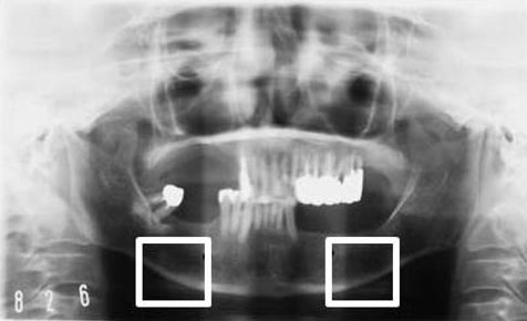
Fig. 1 A panoramic radiograph shows two boxes corresponding to the lower border of the inferior cortex below the mental foramen on the right and left sides of the mandible. Boxes represent the selected region of interest (ROI).

Fig. 2 A second-order polynomial function used to carry out the distance measurements between the boundaries of the cortical bone on the right (A) and the left (B) sides.

Fig. 3 Mandibular cortical bone images show the left cortex affected by noise (boxes in red), also shown in enlarged view. Original images of the left region of interest (A and B), after the high-pass filter (C and D), and after the second order polynomial functions (E and F).

Fig. 4 Histogram-based automatic clustering (HAC) algorithm shows the results of the right mandibular cortical bone. A. Original histogram. B. After border effect removal. C. The most desirable cluster for mandibular cortical width measurement(MCW) (optimized k cluster number 4).
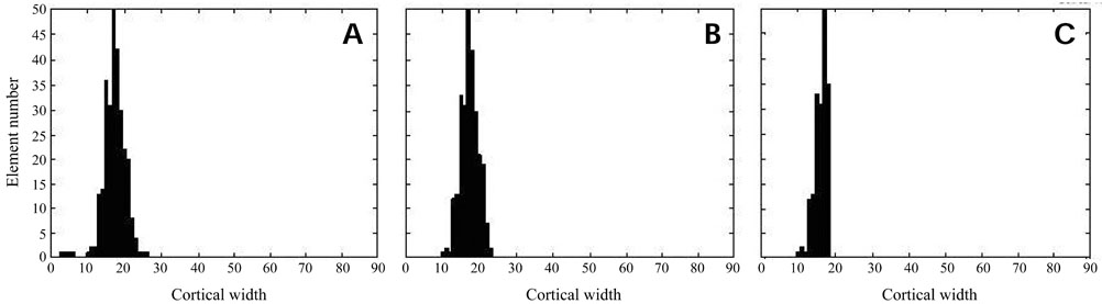
Fig. 5 Histogram-based automatic clustering (HAC) algorithm shows the results of the left mandibular cortical bone. A. Original histogram. B. After border effect removal. C. The most desirable cluster for mandibular cortical width measurement (MCW) (optimized k cluster number 2).
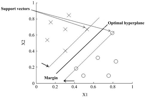
Fig. 6 Support vector machine classification with a linear optimal hyperplane that maximizes the separating margin between the two classes marked as O and X.
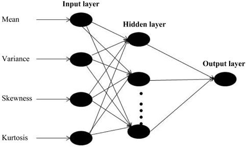
Fig. 7 Structure of typical back propagation neural network model shows four inputs with one hidden layer and one output layer.
Cited by 1 articles
-
IDIOS: An innovative index for evaluating dental imaging-based osteoporosis screening indices
Imad Barngkgei, Esam Halboub, Abeer Abdulkareem Almashraqi, Razan Khattab, Iyad Al Haffar
Imaging Sci Dent. 2016;46(3):185-202. doi: 10.5624/isd.2016.46.3.185.
Reference
-
1. Osteoporosis prevention, diagnosis and therapy. NIH Consensus Statement. 2000. 17:p. 1–45.2. Johnell O, Kanis JA. An estimate of the worldwide prevalence, mortality and disability associated with hip fracture. Osteoporos Int. 2004; 15:897–902.
Article3. Horner K, Devlin H. The relationship between mandibular bone mineral density and panoramic radiographic measurements. J Dent. 1998; 26:337–343.
Article4. Klemetti E, Kolmakov S, Kröger H. Pantomography in assessment of the osteoporosis risk group. Scand J Dent Res. 1994; 102:68–72.
Article5. Hildebolt CF. Osteoporosis and oral bone loss. Dentomaxillofac Radiol. 1997; 26:3–15.
Article6. White SC, Rudolph DJ. Alterations of the trabecular pattern of the jaws in patients with osteoporosis. Oral Surg Oral Med Oral Pathol Oral Radiol Endod. 1999; 88:628–635.
Article7. White SC, Taguchi A, Kao D, Wu S, Service SK, Yoon D, et al. Clinical and panoramic predictors of femur bone mineral density. Osteoporos Int. 2005; 16:339–346.
Article8. Devlin H, Karayianni K, Mitsea A, Jacobs R, Lindh C, van der Stelt P, et al. Diagnosing osteoporosis by using dental panoramic radiographs: the OSTEODENT project. Oral Surg Oral Med Oral Pathol Oral Radiol Endod. 2007; 104:821–828.
Article9. Arifin AZ, Asano A, Taguchi A, Nakamoto T, Ohtsuka M, Tsuda M, et al. Computer-aided system for measuring the mandibular cortical width on dental panoramic radiographs in identifying postmenopausal women with low bone mineral density. Osteoporos Int. 2006; 17:753–759.
Article10. Devlin H, Allen PD, Graham J, Jacobs R, Karayianni K, Lindh C, et al. Automated osteoporosis risk assessment by dentists: a new pathway to diagnosis. Bone. 2007; 40:835–842.
Article11. Allen PD, Graham J, Farnell DJ, Harrison EJ, Jacobs R, Nicopolou-Karayianni K, et al. Detecting reduced bone mineral density from dental radiographs using statistical shape models. IEEE Trans Inf Technol Biomed. 2007; 11:601–610.
Article12. Roberts M, Yuan J, Graham J, Jacobs R, Devlin H. Changes in mandibular cortical width measurements with age in men and women. Osteoporos Int. 2011; 22:1915–1925.
Article13. Kavitha MS, Samopa F, Asano A, Taguchi A, Sanada M. Computer-aided measurement of mandibular cortical width on dental panoramic radiographs for identifying osteoporosis. J Investig Clin Dent. 2012; 3:36–44.
Article14. Nakamoto T, Taguchi A, Ohtsuka M, Suei Y, Fujita M, Tanimoto K, et al. Dental panoramic radiograph as a tool to detect postmenopausal women with low bone mineral density: untrained general dental practitioners' diagnostic performance. Osteoporos Int. 2003; 14:659–664.15. Lee K, Taguchi A, Ishii K, Suei Y, Fujita M, Nakamoto T, et al. Visual assessment of the mandibular cortex on panoramic radiographs to identify postmenopausal women with low bone mineral densities. Oral Surg Oral Med Oral Pathol Oral Radiol Endod. 2005; 100:226–231.
Article16. Taguchi A, Asano A, Ohtsuka M, Nakamoto T, Suei Y, Tsuda M, et al. Observer performance in diagnosing osteoporosis by dental panoramic radiographs: results from the osteoporosis screening project in dentistry (OSPD). Bone. 2008; 43:209–213.
Article17. World Health Organization. Assessment of fracture risk and its application to screening for postmenopausal women osteoporosis. Geneva: World Health Organization;1994.18. Fujiwara S, Kasagi F, Masunari N, Naito K, Suzuki G, Fukunaga M. Fracture prediction from bone mineral density in Japanese men and women. J Bone Miner Res. 2003; 18:1547–1553.
Article19. Orimo H, Hayashi Y, Fukunaga M, Sone T, Fujiwara S, Shiraki M, et al. Diagnostic criteria for primary osteoporosis: year 2000 revision. J Bone Miner Metab. 2001; 19:331–337.
Article20. Gonzalez RC, Woods RE. Digital image processing. Englewood Cliffs: Addison-Wesley Pub;1992.21. Ketchen DJ, Shook CL. The application of cluster analysis in strategic management research: an analysis and critique. Strat Mgmt J. 1996; 17:441–458.22. Manning CD, Raghavan P, Schütze H. Introduction to information retrieval. New York: Cambridge University Press;2008.23. Hartigan JA. Clustering algorithms: probability & mathematical statistics. New York: Wiley;1975.24. Wee LJ, Tan TW, Ranganathan S. SVM-based prediction of caspase substrate cleavage sites. BMC Bioinformatics. 2006; 7:Suppl 5. S14.
Article25. Burges CJ. A tutorial on support vector machines for pattern recognition. Data Min Knowl Discov. 1998; 2:121–167.26. Vapnik VN. The nature of statistical learning theory. New York: Springer;1995.27. Vapnik VN. Statistical learning theory. New York: Wiley;1998.28. Ciesielski K, Sacha JP, Cios K. Synthesis of feedforward networks in supremum error bound. IEEE Trans Neural Netw. 2000; 11:1213–1227.29. Back AD, Chen T. Universal approximation of multiple nonlinear operators by neural networks. Neural Comput. 2002; 14:2561–2566.
Article30. Aleksander I, Morton H. An introduction to neural computing. London: Chapman and Hall;1990.31. Schölkopf B, Smola AJ. Learning with kernels. Cambridge: MIT Press;2002.32. Haykin S. Neural networks: a comprehensive foundation. 2nd ed. Englewood Cliffs: Prentice Hall;1999.33. Karayianni K, Horner K, Mitsea A, Berkas L, Mastoris M, Jacobs R, et al. Accuracy in osteoporosis diagnosis of a combination of mandibular cortical width measurement on dental panoramic radiographs and a clinical risk index (OSIRIS): the OSTEODENT project. Bone. 2007; 40:223–229.
Article34. Tuceryan M, Jain AK. Texture analysis. In : Chen CH, Pau LF, Wang PS, editors. The handbook of pattern recognition and computer vision. Singapore: World Scientific Publishing Co.;1998. p. 207–248.35. Kavitha MS, Asano A, Taguchi A, Kurita T, Sanada M. Diagnosis of osteoporosis from dental panoramic radiographs using the support vector machine method in a computer-aided system. BMC Med Imaging. 2012; 12:1.
Article36. Muramatsu C, Matsumoto T, Hayashi T, Hara T, Katsumata A, Zhou X, et al. Automated measurement of mandibular cortical width on dental panoramic radiographs. Int J Comput Assist Radiol Surg. 2000; (in press).
Article37. Yosue T, Brooks SL. The appearance of the mental foramina on panoramic and periapical radiographs. II. Experimental evaluation. Oral Surg Oral Med Oral Pathol. 1989; 68:488–492.
- Full Text Links
-
- Actions
-
Cited
- CITED
-
- Close
- Share
-
- Similar articles
-
- Texture Analysis of Supraspinatus Ultrasound Image for Computer Aided Diagnostic System
- Improving the Performance of Text Categorization Models used for the Selection of High Quality Articles
- Machine Learning on Early Diagnosis of Depression
- Review of Machine Learning Algorithms for Diagnosing Mental Illness
- A Clustering Tool Using Particle Swarm Optimization for DNA Chip Data
