Disseminated non-Hodgkin's lymphoma presenting as bilateral salivary gland enlargement: a case report
- Affiliations
-
- 1Department of Oral Medicine and Radiology, College of Dental Sciences, Davangere, India. manju4744@yahoo.co.in
- 2Department of Oral Medicine and Radiology, SDM College of Dental Sciences and Hospital, Dharwad, India.
- 3Department of Oral Medicine and Radiology, Jamia Milia Islamia University, New Delhi, India.
- KMID: 2167449
- DOI: http://doi.org/10.5624/isd.2013.43.1.59
Abstract
- Non-Hodgkin's lymphoma (NHL) constitutes a group of malignancies those arises from cellular components of lymphoid or extranodal tissues. The head and neck is the most common area for the presentation of these lymphoproliferative disorders. Primary involvement of salivary glands is uncommon. This report described a case of a 73-year-old female patient who presented with involvement of both nodal and extranodal sites, with predominant involvement of salivary glands. The tumor staging worked up along with imaging, histopathological, and immunohistochemical findings were discussed. Computed tomographic images showed the involvement of Waldeyer's ring, larynx, orbit, and spleen. This report described imaging and prognostic tumor markers in diagnosing, treatment planning, and prognosis.
MeSH Terms
Figure
-
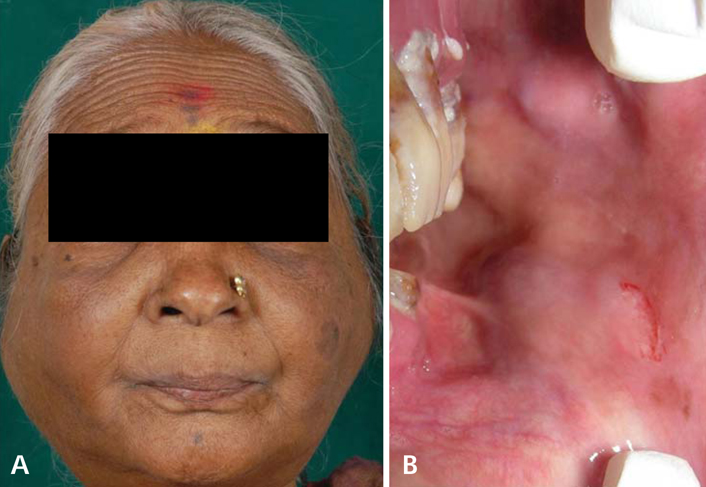
Fig. 1 A. Bilateral enlargements of parotid and submandibular glands are seen. B. Two swellings measuring 2 cm×3 cm are seen in the buccal vestibule in the region of the upper and lower second molar.

Fig. 2 A. A high resolution ultrasonographic image shows the gross enlargement of the right parotid gland measuring 79 mm×31 mm and the involvement of both deep and superficial lobes by hypoechoic areas with a few echogenic septae traversing in between is evident. B. A high resolution ultrasonographic image shows the enlargement of the submandibular glands showing heterogeneous echogenicity with distorted architecture.

Fig. 3 A. Contrast-enhanced computed tomography shows diffuse symmetrical enlargement of the parotid gland with heterogeneous moderate enhancement. B. Contrast-enhanced computed tomography shows diffuse symmetrical enlargement of the submandibular gland with heterogeneous moderate enhancement.

Fig. 4 A. Histopathological examination shows diffuse large B-cells with moderate cytoplasm and large round vesicular nuclei having prominent nucleoli (H&E stain, ×20). B. Immunohistochemical staining shows positive staining for CD20 (IHC, ×40).
Reference
-
1. Urquhart A, Berg R. Hodgkin's and non-Hodgkin's lymphoma of the head and neck. Laryngoscope. 2001. 111:1565–1569.
Article2. van der Waal RI, Huijgens PC, van der Valk P, van der Waal I. Characteristics of 40 primary extranodal non-Hodgkin lymphomas of the oral cavity in perspective of the new WHO classification and the International Prognostic Index. Int J Oral Maxillofac Surg. 2005. 34:391–395.
Article3. Nadendla LK, Meduri V, Paramkusam G. Imaging characteristics of diffuse large cell extra nodal non-Hodgkin's lymphoma involving the palate and maxillary sinus: a case report. Imaging Sci Dent. 2012. 42:111–114.
Article4. Newton R, Ferlay J, Beral V, Devesa SS. The epidemiology of non-Hodgkin's lymphoma: comparison of nodal and extranodal sites. Int J Cancer. 1997. 72:923–930.
Article5. Teh CS, Chong SY. An unusual presentation of lymphoma of the head and neck region. Med J Malaysia. 2011. 66:264–265.6. Zagolski O, Dwivedi R, Kazi R, Subramanian S. Non-Hodgkin's lymphoma of the sino-nasal tract in children. J Cancer Res Ther. 2010. 6:5–10.7. Ioachim HL, Ryan JR, Blaugrund SM. Salivary gland lymph nodes. The site of lymphadenopathies and lymphomas associated with human immunodeficiency virus infection. Arch Pathol Lab Med. 1988. 112:1224–1228.8. Aiken AH, Glastonbury C. Imaging Hodgkin and non-Hodgkin lymphoma in the head and neck. Radiol Clin North Am. 2008. 46:363–378.
Article9. Carbone PP, Kaplan HS, Musshoff K, Smithers DW, Tubiana M. Report of the committee on Hodgkin's disease staging classification. Cancer Res. 1971. 31:1860–1861.10. Burns FM, Parks S, Marley JJ. Primary non-Hodgkin's lymphoma of the mandible manifesting as a dentigerous cyst. Oral Surg. 2011. 4:73–76.
Article11. Gleeson MJ, Bennett MH, Cawson RA. Lymphomas of salivary glands. Cancer. 1986. 58:699–704.
Article12. DePeña CA, Van Tassel P, Lee YY. Lymphoma of the head and neck. Radiol Clin North Am. 1990. 28:723–743.13. Roh JL, Huh J, Suh C. Primary non-Hodgkin's lymphomas of the major salivary glands. J Surg Oncol. 2008. 97:35–39.
Article14. Batsakis JG. Primary lymphomas of the major salivary glands. Ann Otol Rhinol Laryngol. 1986. 95:107–108.
