Reference line-pair values of panoramic radiographs using an arch-form phantom stand to assess clinical image quality
- Affiliations
-
- 1Department of Oral and Maxillofacial Radiology and Dental Research Institute, School of Dentistry, Seoul National University, Seoul, Korea.
- 2Department of Oral and Maxillofacial Radiology, College of Dentistry, Dankook University, Cheonan, Korea.
- 3Department of Oral and Maxillofacial Radiology, BK21 Craniomaxillofacial Life Science, and Dental Research Institute, School of Dentistry, Seoul National University, Seoul, Korea. raylee@snu.ac.kr
- KMID: 2167441
- DOI: http://doi.org/10.5624/isd.2013.43.1.7
Abstract
- PURPOSE
This study was performed to suggest reference line-pair values of panoramic images with clinically desirable qualities using an arch-form phantom stand.
MATERIALS AND METHODS
The line-pair test phantom was chosen. A real skull model was selected for setting the arch-form model of the phantom stand. The phantom stand had slits in four regions (incisor, premolar, molar, TMJ). Four raw images of the test phantom in each region and one raw image of the real skull were converted into 50 test phantom images and 50 skull phantom images with various line-pair values. 50 post-processed real skull phantom images were divided into 4 groups and were randomly submitted to 14 evaluators. Image quality was graded on a 4 point scale (1. good, 2. normal, 3. poor but interpretable, and 4. not interpretable). The reference line pair was determined as the first line-pair value scored less than 2 points. RESULT: The mean scores tended to decrease as the line-pair values increased. The reference line-pair values were 3.19 LP/mm in the incisor, 2.32 LP/mm in the premolar and TMJ, and 1.88 LP/mm in the molar region.
CONCLUSION
Image quality evaluation methods and criteria should be able to assess various regions considering the characteristics of panoramic systems. This study suggested overall and regional reference line-pair values and established a set of standard values for them.
Figure
-
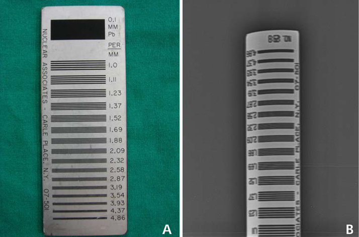
Fig. 1 A. Line-pair test phantom (Nuclear Associates model 07-501 SER. NO.12913). B. The radiographic image of the line-pair test phantom.
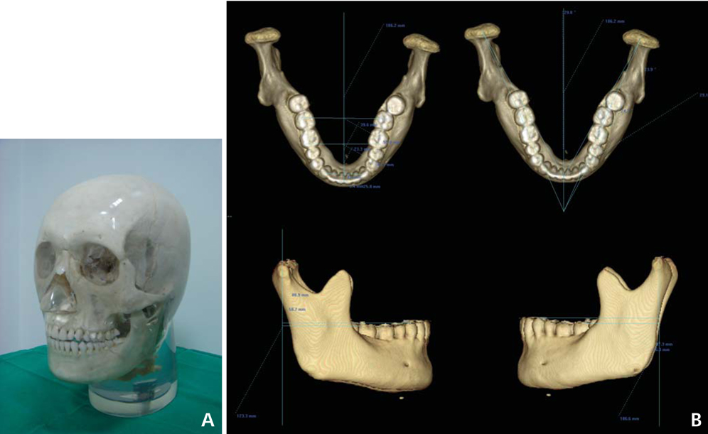
Fig. 2 A. Transparent real skull X-ray phantom. B. The structural connection of the mandibular condyle with the lower dental arch of the skull phantom is measured on the 3D CT images.
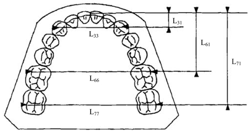
Fig. 3 Reference points and associated measurements. The dimensions of the dental arches are determined according to three sagittal and three transverse measurements. (This image is cited from a study by Raberin et al.8)

Fig. 4 A. The phantom stand: Slits are prepared in 4 regions including the incisor, right premolar, left molar, and right TMJ regions to hold the test phantoms. B. Positioning the phantom stand for panoramic image acquisition using the tripod connection.
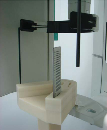
Fig. 5 The selected line-pair phantom is inserted in the right TMJ slit of the phantom stand and these are positioned for panoramic image acquisition.
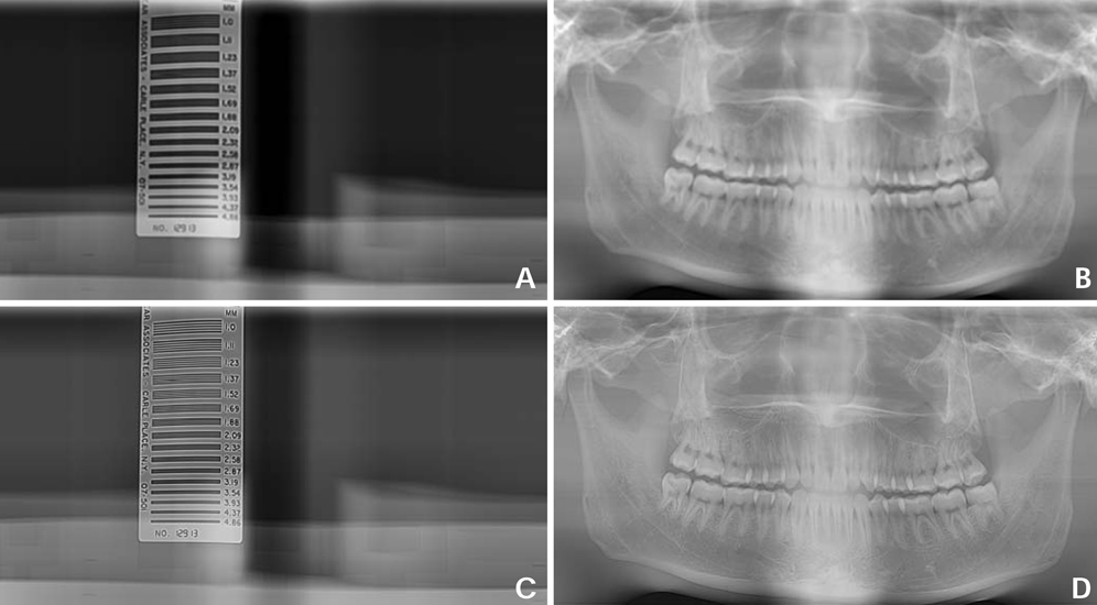
Fig. 6 The post-processed test phantom radiographs and corresponding post-processed real skull phantom radiographs, respectively. A. The post-processed image of the test phantom in premolar region showing a 1.11 line-pair value (1.11 LP/mm). B. The post-processed image of the real skull phantom under the same parameter settings for "A" image post-processing. C. The post-processed image of the test phantom in the premolar region showing a 2.87 line-pair value (2.87 LP/mm). D. The post-processed image of the real skull phantom under the same parameter settings for "C" image post-processing.
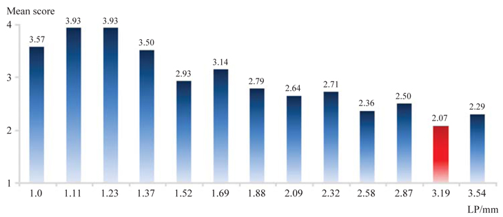
Fig. 7 The mean scores in the incisor group tend to decrease as the line-pair values increased. 3.19 LP/mm is determined to be the reference line-pair value.
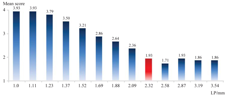
Fig. 8 The mean scores in the premolar group tend to decrease as the line-pair values increases. 2.32 LP/mm is determined to be the reference line-pair value.
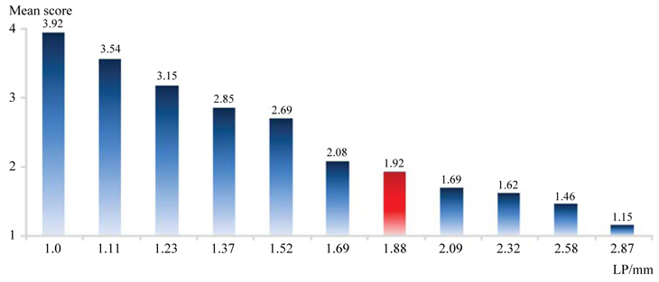
Fig. 9 The mean scores in the molar group consistently decrease as the line-pair values increases. 1.88 LP/mm is determined to be the reference line-pair value.
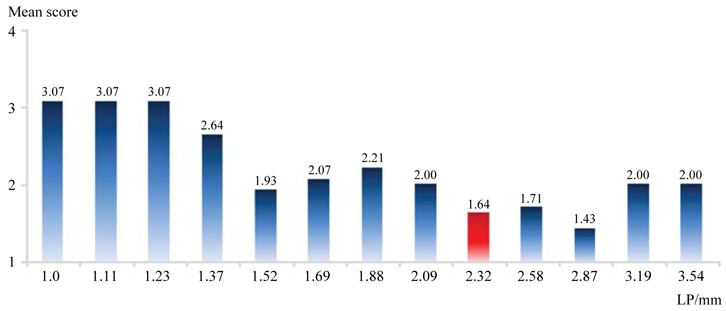
Fig. 10 The mean scores in the TMJ group tend to decrease as the line-pair values increases. 2.32 LP/mm is determined to be the reference line-pair value.
Cited by 1 articles
-
Contrast reference values in panoramic radiographic images using an arch-form phantom stand
Jae-Myung Shin, Chena Lee, Jo-Eun Kim, Kyung-Hoe Huh, Won-Jin Yi, Min-Suk Heo, Soon-Chul Choi, Sam-Sun Lee
Imaging Sci Dent. 2016;46(3):203-210. doi: 10.5624/isd.2016.46.3.203.
Reference
-
1. Yoo S, Kim GY, Hammoud R, Elder E, Pawlicki T, Guan H, et al. A quality assurance program for the on-board imagers. Med Phys. 2006. 33:4431–4447.2. Enforcement regulations of the installation and operation of special medical equipments. Enforcement regulation No.65 of Ministry of Health and Welfare. 2003.3. Lee SH, Choe YH, Chung SY, Kim MH, Kim EK, Oh KK, et al. Establishment of quality assessment standard for mammographic equipments: evaluation of phantom and clinical images. J Korean Radiol Soc. 2005. 53:117–127.
Article4. Korean Institute for Accreditation of Medical Imaging [internet]. Table for mammographic quality control. Available from: http://www.ikiami.or.kr/Data/KMI501QD.aspx.5. Kin HJ, Lee HG, Jeong JB, Im CI, Son HK. A study on guidance for medical radiation safety. Final report. 2010. Seoul: Korea Food and Drug Administration;Report No.: 10171Radiology 457.6. White SC, Pharoah MJ. Oral Radiology: principles and interpretation. 2009. 6th ed. St. Louis: Mosby-Year Book Inc.7. Gopal A, Samant SS. Use of a line-pair resolution phantom for comprehensive quality assurance of electronic portal imaging devices based on fundamental imaging metrics. Med Phys. 2009. 36:2006–2015.
Article8. Raberin M, Laumon B, Martin JL, Brunner F. Dimensions and form of dental arches in subjects with normal occlusions. Am J Orthod Dentofacial Orthop. 1993. 104:67–72.
Article9. DIN German Institute for Standardization. DIN 6868-151. Image quality assurance in diagnostic X-ray departments - Part 151: acceptance testing of dental radiographic equipment accordance to RöV - Rules for the inspection of image quality after installation, maintenance and modification. 2010. Berlin: DIN Deutsches Institut fur Normung e. V..
- Full Text Links
-
- Actions
-
Cited
- CITED
-
- Close
- Share
-
- Similar articles
-
- Contrast reference values in panoramic radiographic images using an arch-form phantom stand
- A study of panoramic focal trough for the six-year-old child
- Development of a new ball-type phantom for evaluation of the image layer of panoramic radiography
- Clinical image quality evaluation for panoramic radiography in Korean dental clinics
- Development of Dual-Window Phantom for Output Measurement of Medical Linacs
