Imaging Sci Dent.
2011 Dec;41(4):177-181. 10.5624/isd.2011.41.4.177.
Pyknodysostosis: report of a rare case with review of literature
- Affiliations
-
- 1Department of Oral Medicine and Radiology, Mar Baselios Dental College, Kothamangalam, India. krksmile@gmail.com
- 2Department of Prosthodontics, Mar Baselios Dental College, Kothamangalam, India.
- KMID: 2167404
- DOI: http://doi.org/10.5624/isd.2011.41.4.177
Abstract
- Pyknodysostosis is a rare autosomal recessive disorder characterized by the post natal onset of short limbs, short stature, and generalized hyperostosis along with acro-osteolysis with sclerosis of the terminal phalanges, a feature that is considered essentially pathognomonic. Other features include persistence of fontanelles, delayed closure of sutures, wormian bones, absence of frontal sinuses, and obtuse mandibular gonial angle with relative mandibular prognathism. We report a case of 17-year-old girl who presented with a chief complaint of retention of deciduous teeth. General physical examination demonstrated short stature, frontal and parietal bossing, depressed nasal bridge, beaked nose, hypoplastic midface, wrinkled skin over the finger tips, and nail abnormalities. Radiographs showed multiple impacted permanent and supernumerary teeth, hypoplastic paranasal sinuses with acro-osteolysis of terminal phalanges, and open fontanelles, and sutures along with wormian bones in the lambdoidal region.
MeSH Terms
Figure
-

Fig. 1 A and B. Clinical photographs show frontal bossing, beaked nose and midfacial hypoplasia.

Fig. 2 Intraoral photograph shows V-shaped high arched palate with midline groove and multiple retained deciduous teeth.
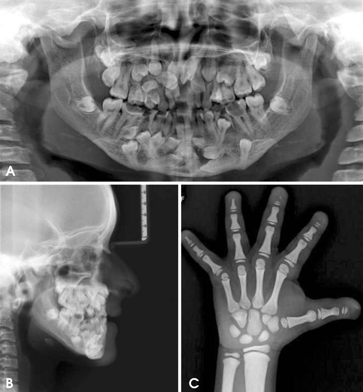
Fig. 3 A. Panoramic radiograph shows multiple impacted permanent and supernumerary teeth. B. Lateral cephalogram shows hypoplastic paranasal sinuses with relative mandibular prognathism. C. Hand-wrist radiograph shows acro-osteolysis of terminal phalanges.

Fig. 4 A and B. Three-dimensional CT images show open fontanelles and sutures, wormian bones in the lambdoidal region.
Reference
-
1. Maroteaux P, Lamy M. Pyknodysostosis. Presse Med. 1962. 70:999–1002.2. Fleming KW, Barest G, Sakai O. Dental and facial bone abnormalities in pyknodysostosis: CT findings. AJNR Am J Neuroradiol. 2007. 28:132–134.3. Mujawar Q, Naganoor R, Patil H, Thobbi AN, Ukkali S, Malagi N. Pyknodysostosis with unusual findings: a case report. Cases J. 2009. 2:6544.4. Moniz N, Queiroz EA, Freitas RR, Felix VB. Mandibular reconstruction with autogenous graft in patient presenting pyknodysostosis: case report. J Oral Maxillofac Surg. 2006. 64:1292–1295.
Article5. Hunt NP, Cunningham SJ, Adnan N, Harris M. The dental, craniofacial, and biochemical features of pyknodysostosis: a report of three new cases. J Oral Maxillofac Surg. 1998. 56:497–504.
Article6. Schmitz JP, Gassmann CJ, Bauer AM, Smith BR. Mandibular reconstruction in a patient with pyknodysostosis. J Oral Maxillofac Surg. 1996. 54:513–517.
Article7. Motyckova G, Fisher DE. Pycnodysostosis: role and regulation of cathepsin K in osteoclast function and human disease. Curr Mol Med. 2002. 2:407–421.
Article8. Elmore SM. Pycnodysostosis: a review. J Bone Joint Surg. 1967. 49:153–162.9. Landa S, Esteban S, Montes E, Santamaria J, Vitoria A, Santolaya JM. Maxillofacial alterations in a family with pycnodysostosis. Med Oral. 2000. 5:169–176.10. Rajendran R. Rajendran R, Sivapathasundharam B, editors. Diseases of bone and joints. Shafer's textbook of oral pathology. 2010. 6th ed. Noida: Elsevier;701.11. Soares LF, Souza I, Cardoso AS, Pomarico L. Pyknodysostosis: oral findings and differential diagnosis. J Indian Soc Pedod Prev Dent. 2008. 26:S23–S25.
- Full Text Links
-
- Actions
-
Cited
- CITED
-
- Close
- Share
-
- Similar articles
-
- Posterior Epidural Migration of a Sequestrated Intervertebral Lumbar Disc: A Case Report
- Cystic Lymphangioma of Rectum-A Case Report and Review of Literature
- Bilateral Clavicular Fracture Related to Birth in Newborn: A Case Report with Literature Review
- Peripheral Cerebral Arterial Aneurysm: Case Report and Review of the Literature
- Eccrine Nevus on the Neck: A Case Report and Review of the Literature
