Expression of EGFR and Microvessel Density in Middle Ear Cholesteatoma
- Affiliations
-
- 1Department of Otolaryngology-Head and Neck Surgery, Hanyang University College of Medicine, Seoul, Korea. shleemd@hanyang.ac.kr
Abstract
OBJECTIVES
Cholesteatoma destructs bony tissue by the interactions between hyperproliferative epithelial cells and subepithelial inflammatory cells. The aim of this study was to evaluate the expression of epidermal growth factor receptor (EGFR) and microvessel density (MVD) in middle ear cholesteatoma tissue in an effort to determine the relationship between expression of EGFR and neovascularization.
METHODS
We evaluated the expression of EGFR and MVD by immunohistochemical staining for CD31 and Factor VIII in 32 cholesteatoma tissue samples and 7 normal postauricular skin samples. We also analyzed the correlation between EGFR expression and MVD.
RESULTS
The expression of EGFR was higher in cholesteatoma than in postauricular skin, but the difference was not statistically significant. EGFR was more highly expressed in the suprabasal layer than in the basal layer. Using CD31 and Factor VIII, we analyzed the MVD and found that it was significantly higher in cholesteatoma than in postauricular skin, and significantly correlated with the expression of EGFR.
CONCLUSION
Our results suggest that overexpression of EGFR and neovascularization are correlated with the growth of cholesteatoma.
MeSH Terms
Figure
-

Fig. 1 Immunohistochemical demonstration of epidermal growth factor receptor in cholesteatoma (×400). Negative (A), weakly positive (B), moderately positive (C) and strongly positive (D) findings.
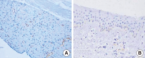
Fig. 2 Immunohistochemical demonstration of microvessel density using anti-CD31 antibody in a cholesteatoma (A) and postauricular skin (B) (×200). Cholesteatoma tissue shows high microvessel density, but postauricular skin shows low microvessel density.
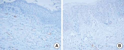
Fig. 3 Immunohistochemical demonstration of microvessel density using anti-Factor VIII antibody in a cholesteatoma (A) and postauricular skin (B) (×200). Cholesteatoma tissue shows high microvessel density, but postauricular skin shows low microvessel density.
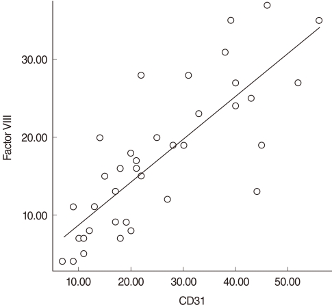
Fig. 4 Correlation between CD31 and Factor VIII. A positive correlation between CD31 and Factor VIII was identified. The correlation efficient was 0.814 (by Spearman correlation test) with a P<0.001.
Reference
-
1. Marenda SA, Aufdemorte TB. Localization of cytokines in cholesteatoma tissue. Otolaryngol Head Neck Surg. 1995; 3. 112(3):359–368. PMID: 7532849.
Article2. Buckingham RA. Etiology of a middle ear cholesteatoma, Kodachrome study of pathogenesis. Ann Otol Rhinol Laryngol. 1968; 12. 77(6):1054–1058. PMID: 5724411.3. Downward J, Yarden Y, Mayes E, Scrace G, Totty N, Stockwell P, et al. Close similarity of epidermal growth factor receptor and v-erb-B oncogene protein sequences. Nature. 1984; 2. 9-15. 307(5951):521–527. PMID: 6320011.
Article4. Mor F, Quintana FJ, Cohen IR. Angiogenesis-inflammation cross-talk: vascular endothelial growth factor is secreted by activated T cells and induces Th1 polarization. J Immunol. 2004; 4. 01. 172(7):4618–4623. PMID: 15034080.
Article5. Folkman J, Watson K, Ingber D, Hanahan D. Induction of angiogenesis during the transition from hyperplasia to neoplasia. Nature. 1989; 5. 04. 339(6219):58–61. PMID: 2469964.6. Bujia J, Holly A, Stammberger M, Sudhoff H. Angiogenesis in cholesteatoma of the middle ear. Acta Otorrinolaringol Esp. 1996; May-Jun. 47(3):187–192. PMID: 8924281.7. Sudhoff H, Dazert S, Gonzales AM, Borkowski G, Park SY, Baird A, et al. Angiogenesis and angiogenic growth factors in middle ear cholesteatoma. Am J Otol. 2000; 11. 21(6):793–798. PMID: 11078065.8. Shinoda H, Huang CC. Expressions of c-jun and p53 proteins in human middle ear cholesteatoma: relationship to keratinocyte proliferation, differentiation, and programmed cell death. Laryngoscope. 1995; 11. 105(11):1232–1237. PMID: 7475882.
Article9. Costa S, Stamm H, Almendral A, Ludwig H, Wyss R, Fabbro D, et al. Predictive value of EGF receptor in breast cancer. Lancet. 1988; 11. 26. 2(8622):1258. PMID: 2903994.
Article10. Xian W, Kiguchi K, Imamoto A, Rupp T, Zilberstein A, DiGiovanni J. Activation of the epidermal growth factor receptor by skin tumor promoters and in skin tumors from SENCAR mice. Cell Growth Differ. 1995; 11. 6(11):1447–1455. PMID: 8562483.11. Fleming TP, Saxena A, Clark WC, Robertson JT, Oldfield EH, Aaronson SA, et al. Amplification and/or overexpression of platelet-derived growth factor receptors and epidermal growth factor receptor in human glial tumors. Cancer Res. 1992; 8. 15. 52(16):4550–4553. PMID: 1322795.12. Santini J, Formento JL, Francoual M, Milano G, Schneider M, Dassonville O, et al. Characterization, quantification, and potential clinical value of the epidermal growth factor receptor in head and neck squamous cell carcinomas. Head Neck. 1991; Mar-Apr. 13(2):132–139. PMID: 2022478.
Article13. Bujia J, Kim C, Holly A, Sudhoff H, Ostos P, Kastenbauer E. Epidermal growth factor receptor (EGF-R) in human middle ear cholesteatoma: an analysis of protein production and gene expression. Am J Otol. 1996; 3. 17(2):203–206. PMID: 8723947.14. Kim JH, Kim HJ, Park JY. Expressions of epidermal growth factor receptor in the epithelium and cultured keratinocyte of aural cholesteatoma. Korean J Otolaryngol-Head Neck Surg. 1998; 5. 41(5):559–566.15. Ahn JM, Huang CC, Abramson M. Third place--Resident Basic Science Award 1990: Interleukin 1 causing bone destruction in middle ear cholesteatoma. Otolaryngol Head Neck Surg. 1990; 10. 103(4):527–536. PMID: 2174138.16. Schilling V, Bujía J, Negri B, Schulz P, Kastenbauer E. Immunologically activated cells in aural cholesteatoma. Am J Otolaryngol. 1991; Sep-Oct. 12(5):249–253. PMID: 1811419.
Article17. Stammberger M, Bujía J, Schulz P. Correlation of vascular morphology with clinical types in cholesteatoma of the middle ear. Am J Otol. 1994; 5. 15(3):380–382. PMID: 8579144.18. Murray JD, Carlson GW, McLaughlin K, Pennington M, Lynn M, DeRose PB, et al. Tumor angiogenesis as a prognostic factor in laryngeal cancer. Am J Surg. 1997; 11. 174(5):523–526. PMID: 9374229.19. Anniko M, Mendel L. Cholesteatoma: a clinical and morphological analysis. Acta Otolaryngol. 1981; Mar-Apr. 91(3-4):275–283. PMID: 7257760.20. Stammberger M, Holly A, Bujia J. Immunohistochemical studies of vascularization of cholesteatoma. Laryngorhinootologie. 1993; 4. 72(4):170–173. PMID: 7684226.21. Sudhoff H, Fisseler-Eckhoff A, Borkowski G, Luckhaupt H, Stark T, Hildmann H. Sanna M, editor. Cholesteatoma and angiogenesis. Cholesteatoma and mastoid surgery (Proceedings of the Fifth International Conference on Cholesteatoma and Mastoid Surgery). 1997. Roma: CIC Edizioni Internazionali;p. 264–272.
- Full Text Links
-
- Actions
-
Cited
- CITED
-
- Close
- Share
-
- Similar articles
-
- Expressions of Epidermal Growth Factor Receptor in the Epithelium and Cultured Keratinocyte of Aural Cholesteatoma
- Clinical Significance of Expression of Epidermal Growth Factor, Epidermal Growth Factor Receptor, Transforming Growth Factor-alpha in Aural Cholesteatoma
- A Retroauricular Cholesteatoma in Soft Tissue after Tympanomastoidectomy
- Changes of Apoptosis according to the Site and Clinical Stage of Experimental Cholesteatoma
- Immunohistochemical demonstration of Langerhans' cells in middle ear cholesteatoma
