Venous Hemangioma of Parapharyngeal Space with Calcification
- Affiliations
-
- 1Department of Otorhinolaryngology-Head and Neck Surgery, The Catholic University of Korea College of Medicine, Seoul, Korea. hnsdi@catholic.ac.kr
Abstract
- A hemangioma of the parapharyngeal space (PPS) is an extremely rare tumor and is responsible for 0.5-1% of all tumors occurring in the PPS. We report a case of PPS venous hemangioma in a 49-year-old woman presenting with diffuse swelling in the submandibular region. A preoperative computed tomography (CT) scan showed a cystic mass with multiple calcifications in the PPS. The calcific nodules were round and about 2 mm in diameter. The hemangioma was completely resected via a transcervical approach. During surgery, we found several calcific nodules, which represented phleoboliths or areas of thrombosis with dystrophic calcification. Despite its rarity, a venous hemangioma of the PPS should be considered in a differential diagnosis when a cystic mass with calcification is found by CT scan. To our knowledge, this is the first reported case of a PPS venous hemangioma; we describe its pathognomonic findings on imaging.
Keyword
Figure
-
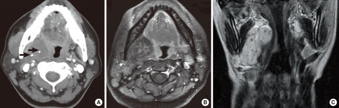
Fig. 1 (A) Contrast-enhanced axial computed tomography scan shows a well-defined and hypoattenuating lesion, 4×3 cm in size, with two spotty calcifications (arrows), on the right parapharyngeal space. (B) The lesion shows low signal intensity on contrast-enhanced axial T1-weighted magnetic resonance images, and the three central areas of low signal intensity with peripheral rim enhancement correspond to the calcifications. (C) T2-weighted coronal magnetic resonance imaging shows that this lesion is hyperintense, making it easier to delineate from adjacent normal soft tissue.
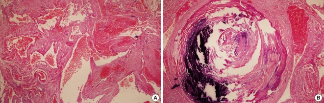
Fig. 2 (A) Microscopic examination revealed that dilated, thick-walled blood vessels were scattered, and some of these vessel were thrombosed (H&E, ×100). (B) A thick muscular-walled vessel containing calcification and thrombus (H&E, ×200).
Reference
-
1. Chrzanowski DS, Powers CN, Reiter ER. Parapharyngeal space hemangioma in a pediatric patient. Otolaryngol Head Neck Surg. 2005; 9. 133(3):455–457. PMID: 16143201.
Article2. Aspestrand F, Kolbenstvedt A. Vascular mass lesions and hypervascular tumors in the head and neck: characteristics at CT, MR imaging and angiography. Acta Radiol. 1995; 3. 36(2):136–141. PMID: 7710791.3. Cankaya H, Unal O, Ugras S, Yuca K, Kiris M. Hemangioma with phleboliths in the sublingual gland: as a cause of submental opacity. Tohoku J Exp Med. 2003; 3. 199(3):187–191. PMID: 12703663.4. Odabasi AO, Metin KK, Mutlu C, Basak S, Erpek G. Intramuscular hemangioma of the masseter muscle. Eur Arch Otorhinolaryngol. 1999; 256(7):366–369. PMID: 10473832.
Article5. Carter LC, Uthman A, Drinnan AJ, Loree TR. Diagnostic dilemma involving calcification in the parapharyngeal space: metastatic thyroid carcinoma masquerading as a deep lobe parotid mass. Oral Surg Oral Med Oral Pathol Oral Radiol Endod. 1997; 12. 84(6):697–702. PMID: 9431542.6. Murata Y, Yamada I, Umehara I, Ishii Y, Okada N. Perfusion and blood-pool scintigraphy in the evaluation of head and neck hemangiomas. J Nucl Med. 1997; 6. 38(6):882–885. PMID: 9189134.7. McMenamin M, Quinn A, Barry H, Sleeman D, Wilson G, Toner M. Cavernous hemangioma in the submandibular gland masquerading as sialadenitis: case report. Oral Surg Oral Med Oral Pathol Oral Radiol Endod. 1997; 8. 84(2):146–148. PMID: 9269015.
- Full Text Links
-
- Actions
-
Cited
- CITED
-
- Close
- Share
-
- Similar articles
-
- A Case of Cavernous Hemangioma in the Parapharyngeal Space as a Neurogenic Tumor
- A Case of Rhabdomyosarcoma in Parapharyngeal Space
- Case Report: A Plunging Ranula Extended into the Parapharyngeal Space
- Two Cases of Wooden Foreign Body in Parapharyngeal Space after Penetrating Injury
- Diagnostic and Surgical Approaches of the Parapharyngeal Space Tumors
