Normal Mastoid Air Cell System Geometry: Has Surface Area Been Overestimated?
- Affiliations
-
- 1Department of Otorhinolaryngology-Head and Neck Surgery, Ewha Womans University School of Medicine, Seoul, Korea. byunsw@ewha.ac.kr
- 2Department of Radiology, Ewha Womans University School of Medicine, Seoul, Korea.
- KMID: 2166280
- DOI: http://doi.org/10.21053/ceo.2016.9.1.27
Abstract
OBJECTIVES
The aim of this study was to emphasize the necessity of a standard in segmentation threshold and algorithm for measuring volume and surface area of mastoid air cell system (MACS).
METHODS
First, we obtained axial computed tomography scans of 54 normal temporal bones from 27 subjects. Then, we manipulated Hounsfield units (HU) image data in DICOM (digital imaging and communications in medicine) files directly using our program. The volume and surface area of MACS were computed and compared at segmentation thresholds (HU) from -700 to 0 at intervals of 50 using 2 algorithms; square pixel based (SP) algorithm and marching square (MS) algorithm.
RESULTS
No significant difference was found between the volumes computed by SP and MS algorithms at each segmentation threshold. The surface area computed by SP algorithm, however, was significantly larger than that by MS algorithm. We could minimize this significant difference through a modification of the SP algorithm. As the lower HU threshold value was set, the smaller volume was measured. The surface area showed a plateau at a threshold of approximately -200 HU. The segmentation threshold had greater influence on the measured volume of MACS than the algorithm did.
CONCLUSION
A standard method for measuring volume and surface area of MACS is thought to be necessary. We suggest that the MS algorithm and -200 HU of the threshold could be a standard in the measurement of volume and surface area of MACS.
MeSH Terms
Figure
-
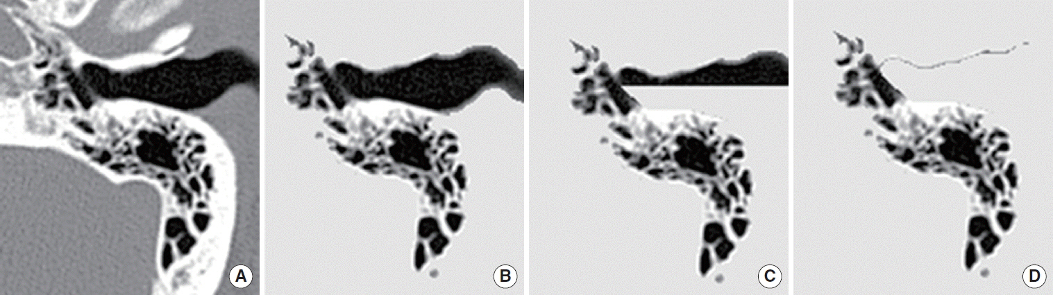
Fig. 1. Grayscale representation (window center, 250; window width, 2,500) of Hounsfield units (HU)-based flood-fill processing. (A, B) Click-driven protected flood-fill safeguards each tentative air pixel (HU<0) and its surrounding pixels within 2-pixel distance. (C, D) Guided flood-fill protects air pixels of one (inner here) side of a given line segment (eardrum here).

Fig. 2. Examples of model extraction by the 2 algorithms (SP, MS) with given segmentation thresholds (HU=–200 and –400 here). HU, Hounsfield units; SP, square pixel based; MS, marching square.

Fig. 3. Schematic illustrations of square pixel based (SP) and marching square (MS) algorithm. In the SP algorithm, each square (pixel) has a center point with Hounsfield units (HU) value, while each square is determined by 4 corner points with HU value in MS algorithm. There are 16 possible configurations of each square in the MS algorithm.

Fig. 4. A modified square pixel (SP) based algorithm to minimize predicted systematic error, which overestimates the surface area of mastoid air cell system. Accordingly the marching square (MS) algorithm is used for computation of both volume and surface area, while the original and the modified algorithms are necessary for volume and surface area, respectively, in the SP based algorithm.

Fig. 5. Volume of mastoid air cell system as calculated by 2 algorithms at a given segmentation threshold from –700 to 0 at intervals of 50. None of the differences of volume at each threshold is statistically significant. Standard errors are drawn to one direction for display simplicity. HU, Hounsfield units.

Fig. 6. Surface area of mastoid air cell system by the 2 algorithms with given segmentation thresholds from –700 to 0 at intervals of 50. In addition, the resulting modification (to avoid systematic error) of square pixel (SP) based algorithm is displayed here, and it is explained in the discussion section. HU, Hounsfield units.

Fig. 7. An explanation for the plateau on the surface area graph. In an example of a square which consists of 4 corner pixels having HU of –500, –180, –220, and +20, respectively, the higher the segmentation threshold is set (HU=–400, –200, or –100), the larger the gray area (air area in 2D slice, volume in 3D MACS) will be measured. However, the length of solid line segment (line segment in 2D slice, surface area in 3D MACS) will be the maximum in HU threshold of –200 in this example. HU, Hounsfield units; MACS, mastoid air cell system; 2D, 2-dimensional; 3D, 3-dimensional.
Reference
-
1. Cinamon U, Sade J. Mastoid and tympanic membrane as pressure buffers: a quantitative study in a middle ear cleft model. Otol Neurotol. 2003; Nov. 24(6):839–42.
Article2. Sade J, Fuchs C. A comparison of mastoid pneumatization in adults and children with cholesteatoma. Eur Arch Otorhinolaryngol. 1994; 251(4):191–5.
Article3. Lesinskas E. Factors affecting the results of nonsurgical treatment of secretory otitis media in adults. Auris Nasus Larynx. 2003; Feb. 30(1):7–14.
Article4. Isono M, Murata K, Azuma H, Ishikawa M, Ito A. Computerized assessment of the mastoid air cell system. Auris Nasus Larynx. 1999; Apr. 26(2):139–45.
Article5. Sirikci A, Bayazit YA, Kervancioglu S, Ozer E, Kanlikama M, Bayram M. Assessment of mastoid air cell size versus sigmoid sinus variables with a tomography-assisted digital image processing program and morphometry. Surg Radiol Anat. 2004; Apr. 26(2):145–8.6. Park MS, Yoo SH, Lee DH. Measurement of surface area in human mastoid air cell system. J Laryngol Otol. 2000; Feb. 114(2):93–6.
Article7. Pata YS, Akbas Y, Unal M, Duce MN, Akbas T, Micozkadioglu D. The relationship between presbycusis and mastoid pneumatization. Yonsei Med J. 2004; Feb. 45(1):68–72.
Article8. Lee DH, Jun BC, Kim DG, Jung MK, Yeo SW. Volume variation of mastoid pneumatization in different age groups: a study by three-dimensional reconstruction based on computed tomography images. Surg Radiol Anat. 2005; Mar. 27(1):37–42.
Article9. Koc A, Ekinci G, Bilgili AM, Akpinar IN, Yakut H, Han T. Evaluation of the mastoid air cell system by high resolution computed tomography: three-dimensional multiplanar volume rendering technique. J Laryngol Otol. 2003; Aug. 117(8):595–8.10. Valtonen HJ, Dietz A, Qvarnberg YH, Nuutinen J. Development of mastoid air cell system in children treated with ventilation tubes for early-onset otitis media: a prospective radiographic 5-year follow-up study. Laryngoscope. 2005; Feb. 115(2):268–73.
Article11. Ahn JY, Park HJ, Park GH, Jeong YS, Kwak HB, Lee YJ, et al. Tympanometry and CT measurement of middle ear volumes in patients with unilateral chronic otitis media. Clin Exp Otorhinolaryngol. 2008; Sep. 1(3):139–42.
Article12. Lee DH, Jun BC, Cho JE, Kim DG, Cho KJ, Yeo SW. Development of mastoid air cell system in Korean normal population: three-dimensional reconstruction based on images from computed tomography. Korean J Otolaryngol-Head Neck Surg. 2004; Jul. 47(7):612–6.13. Python Software Foundation [Internet]. Wilmington (DE): Python Software Foundation;2014 [cited 2014 Dec 1]. Available from: http://www.python.org/.
- Full Text Links
-
- Actions
-
Cited
- CITED
-
- Close
- Share
-
- Similar articles
-
- Development of Mastoid Air Cell System in Korean Normal Population: Three-Dimensional Reconstruction Based on Images from Computed Tomography
- Superior Canal Dehiscence Patients Have Smaller Mastoid Volume than Age- and Sex-Matched Otosclerosis and Temporal Bone Fracture Patients
- Changes of Mastoid Gas Physiology in Virtual Mastoidectomy Model
- Comparison of mastoid air cell volume in patients with or without a pneumatized articular tubercle
- Congenital Cholesteatoma Localized to the Tip of the Mastoid Bone: A Case Report and Possible Etiology
