Effects of Custom-Made Rigid Foot Orthosis on Pes Planus in Children Over 6 Years Old
- Affiliations
-
- 1Department of Rehabilitation Medicine, Chungnam National University Hospital, Daejeon, Korea. asyoung@cnuh.co.kr
- KMID: 2165755
- DOI: http://doi.org/10.5535/arm.2014.38.3.369
Abstract
OBJECTIVE
To identify the effects of a custom-made rigid foot orthosis (RFO) in children over six years old with pes planus.
METHODS
The medical records of 39 children (mean age, 10.3+/-4.09 years) diagnosed with pes planus, fitted with RFOs, and had who more than two consecutive radiological studies were reviewed. The resting calcaneal stance position (RCSP), anteroposterior talocalcaneal angle (APTCA), lateral talocalcaneal angle (LTTCA), the lateral talometatarsal angle (LTTMA), and calcaneal pitch (CP) of both feet were measured to evaluate foot alignment. After diagnosis, children were fitted with a pair of RFOs and recommended to walk with heel strike and reciprocal arm swing to normalize the gait pattern. A follow-up clinical evaluation with radiological measurements was performed after 12-18 months and after 24 months of RFO application. Post-hoc analysis was used to test for significant differences between the radiological indicators and RCSP.
RESULTS
With RFOs, all radiological indicators changed in the corrective direction except LTTCA. RCSP and CP in the third measurement showed significant improvement in comparison with the second and baseline measurements. Additionally, APTCA and LTTMA revealed improvements at the third measurement versus the baseline measurements.
CONCLUSION
This study revealed that radiological indicators improved significantly after 24 months of RFO application. A prospective long-term controlled study with radiographical evaluation is necessary to confirm the therapeutic effects of RFOs and to determine the optimal duration of wear in children with pes planus.
Keyword
MeSH Terms
Figure
-
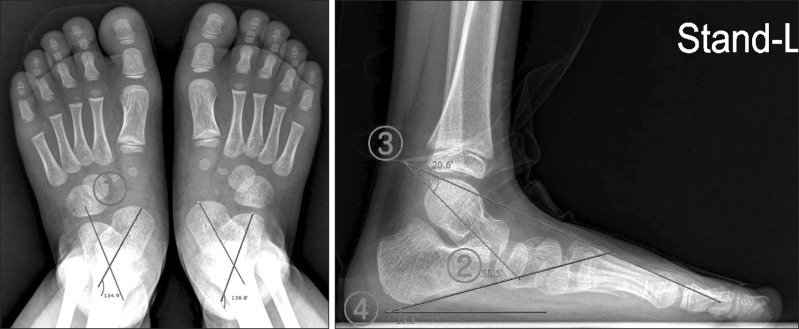
Fig. 1 Anterioposterior view and lateral view of both feet. ①, Anteroposterior talocalcaneal angle; ②, lateral talocalcaneal angle; ③, lateral talometatarsal angle; ④, calcaneal pitch.
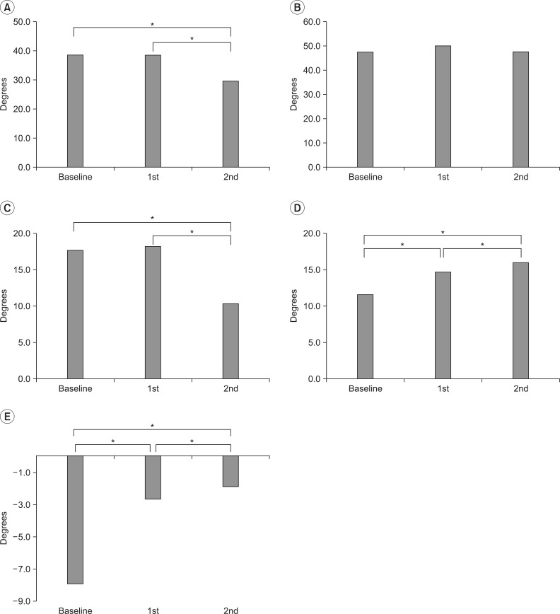
Fig. 2 Serial change in values of anteroposterior talocalcaneal angle (APTCA, A), lateral talocalcaneal angle (LTTCA, B), lateral talometatarsal angle (LTTMA, C), calcaneal pitch (CP, D), and resting calcaneal stance position (RCSP, E).*p<0.05.
Cited by 6 articles
-
Effect of Pressure Based Customized 3-Dimensional Printing Insole in Pediatric Flexible Flat Foot Patients
Si-Wook Lee, Jung-Hoon Choi, Hyuk-Jun Kwon, Kwang-Soon Song
J Korean Foot Ankle Soc. 2020;24(3):113-119. doi: 10.14193/jkfas.2020.24.3.113.Management of Flexible Flatfoot in Chidren and Adolescent
Sun Young Joo, Jung Ryul Kim
J Korean Orthop Assoc. 2016;51(2):109-116. doi: 10.4055/jkoa.2016.51.2.109.Effect of Custom-Molded Foot Orthoses on Foot Pain and Balance in Children With Symptomatic Flexible Flat Feet
Hong-Jae Lee, Kil-Byung Lim, JeeHyun Yoo, Sung-Won Yoon, Hyun-Ju Yun, Tae-Ho Jeong
Ann Rehabil Med. 2015;39(6):905-913. doi: 10.5535/arm.2015.39.6.905.Long-Term Effect of Rigid Foot Orthosis in Children Older Than Six Years With Flexible Flat Foot
Kyo-Jun Youn, So Young Ahn, Bong-Ok Kim, In Sik Park, Soo-Kyung Bok
Ann Rehabil Med. 2019;43(2):224-229. doi: 10.5535/arm.2019.43.2.224.Effect of Foot Orthoses in Children With Symptomatic Flexible Flatfoot Based on Ultrasonography of the Ankle Invertor and Evertor Muscles
Dong Joon Cho, So Young Ahn, Soo-Kyung Bok
Ann Rehabil Med. 2021;45(6):459-470. doi: 10.5535/arm.21137.Change in Plantar Pressure and Plain Radiography in Pediatric Flexible Flatfoot: A Retrospective Cohort Study
Sungjoon Kim, Yong Gyun Kim, Jun Yup Kim, Si-Bog Park, Kyu Hoon Lee
Ann Rehabil Med. 2024;48(5):352-359. doi: 10.5535/arm.240041.
Reference
-
1. Volpon JB. Footprint analysis during the growth period. J Pediatr Orthop. 1994; 14:83–85. PMID: 8113378.
Article2. Pfeiffer M, Kotz R, Ledl T, Hauser G, Sluga M. Prevalence of flat foot in preschool-aged children. Pediatrics. 2006; 118:634–639. PMID: 16882817.
Article3. Jay RM, Schoenhaus HD, Seymour C, Gamble S. The Dynamic Stabilizing Innersole System (DSIS): the management of hyperpronation in children. J Foot Ankle Surg. 1995; 34:124–131. PMID: 7599609.
Article4. Otman S, Basgoze O, Gokce-Kutsal Y. Energy cost of walking with flat feet. Prosthet Orthot Int. 1988; 12:73–76. PMID: 3174409.
Article5. Evans AM, Rome K. A Cochrane review of the evidence for non-surgical interventions for flexible pediatric flat feet. Eur J Phys Rehabil Med. 2011; 47:69–89. PMID: 21448121.6. Whitford D, Esterman A. A randomized controlled trial of two types of in-shoe orthoses in children with flexible excess pronation of the feet. Foot Ankle Int. 2007; 28:715–723. PMID: 17592702.
Article7. Wenger DR, Mauldin D, Speck G, Morgan D, Lieber RL. Corrective shoes and inserts as treatment for flexible flatfoot in infants and children. J Bone Joint Surg Am. 1989; 71:800–810. PMID: 2663868.
Article8. Blake RL, Ferguson H. Foot orthosis for the severe flatfoot in sports. J Am Podiatr Med Assoc. 1991; 81:549–555. PMID: 1774642.
Article9. Blake RL. Inverted functional orthosis. J Am Podiatr Med Assoc. 1986; 76:275–276. PMID: 3712254.
Article
- Full Text Links
-
- Actions
-
Cited
- CITED
-
- Close
- Share
-
- Similar articles
-
- Long-Term Effect of Rigid Foot Orthosis in Children Older Than Six Years With Flexible Flat Foot
- Change of Radiologic Indicators during Putting Foot Orthosis on Flatfoot in Children with Cerebral Palsy
- Pedobarographic Analysis in Functional Foot Orthosis
- Adult-Onset Primary Focal Foot Dystonia Improved with Custom Made Ankle-Foot Orthosis
- Effect of Custom-Molded Foot Orthoses on Foot Pain and Balance in Children With Symptomatic Flexible Flat Feet
