Long-Term Effect of Rigid Foot Orthosis in Children Older Than Six Years With Flexible Flat Foot
- Affiliations
-
- 1Department of Rehabilitation Medicine, Chungnam National University Hospital, Daejeon, Korea. skbok@cnuh.co.kr
- 2Korea Worker's Compensation & Welfare Service Daegu Hospital, Daegu, Korea.
- 3Korean Pedorthic Institute, Goyang, Korea.
- KMID: 2449005
- DOI: http://doi.org/10.5535/arm.2019.43.2.224
Abstract
OBJECTIVE
To evaluate the long-term effect of a custom-made rigid foot orthosis (RFO) in children older than 6 years with pes planus (flat foot).
METHODS
Medical records of 42 children diagnosed with flexible pes planus who were fitted with RFOs based on the inverted technique and underwent more than four consecutive radiological studies were reviewed. Resting calcaneal stance position (RCSP), anteroposterior talocalcaneal angle, lateral talocalcaneal angle, lateral talometatarsal angle, and calcaneal pitch were initially measured in both feet to evaluate alignment. Followup clinical and radiological evaluations were then performed at 12-18, 24-30, 36-42, and ≥48 months after RFO application. Repeated measures analysis of variance was used to evaluate significant differences.
RESULTS
Significant improvements in all radiological indicators and significant progression of RCSP toward the corrective direction were observed after RFO application relative to baseline measurements.
CONCLUSION
According to our findings, RFO can induce significant improvements in calcaneus-related radiographic indices and subsequently improve talus-related radiologic indices.
Keyword
Figure
-
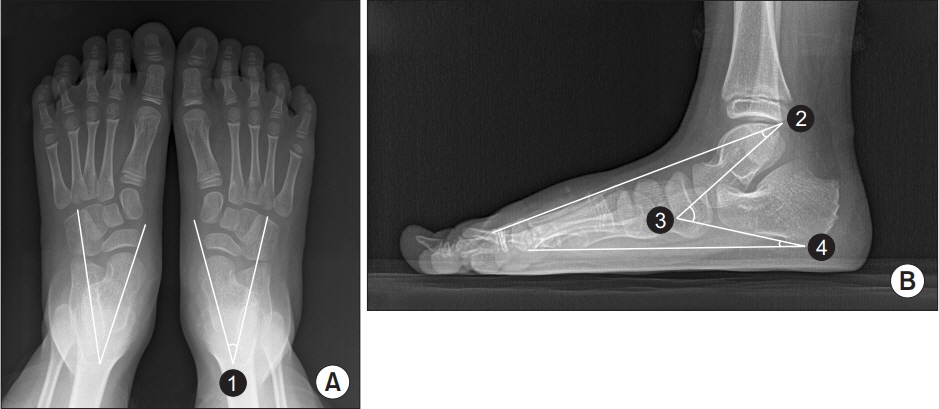
Fig. 1. Representative radiographic images of both feet: (A) anteroposterior view and (B) lateral view indicating ① anteroposterior talocalcaneal angle, ② lateral talometatarsal angle, ③ lateral talocalcaneal angle, and ④ calcaneal pitch.

Fig. 2. Serial changes in anteroposterior talocalcaneal angle (APTCA), lateral talocalcaneal angle (LTTCA), lateral talometatarsal angle (LTTMA), calcaneal pitch (CP), and resting calcaneal stance position (RCSP) over time. *p<0.05; 2nd, 3rd, 4th, and 5th correspond to evaluations conducted at 12–18, 24–30, 36–42, and ≥48 months after rigid foot orthosis application (i.e., baseline).
Cited by 4 articles
-
Flat Foot and Postural Harmony in 6-Year-Old Caucasians: What is Their Relationship?
Teresa Paolucci, Letizia Pezzi, Alice Mannocci, Giuseppe La Torre, Rosa Grazia Bellomo, Raoul Saggini
Ann Rehabil Med. 2020;44(4):320-326. doi: 10.5535/arm.19091.Relationships Between Relative Ankle Muscle Ratios, Severity of Symptoms, and Radiologic Parameters in Adolescent Patients With Symptomatic Flexible Flat Feet
Youngju Shin, So Young Ahn, Soo-Kyung Bok
Ann Rehabil Med. 2021;45(2):123-130. doi: 10.5535/arm.20174.Effect of Foot Orthoses in Children With Symptomatic Flexible Flatfoot Based on Ultrasonography of the Ankle Invertor and Evertor Muscles
Dong Joon Cho, So Young Ahn, Soo-Kyung Bok
Ann Rehabil Med. 2021;45(6):459-470. doi: 10.5535/arm.21137.Change in Plantar Pressure and Plain Radiography in Pediatric Flexible Flatfoot: A Retrospective Cohort Study
Sungjoon Kim, Yong Gyun Kim, Jun Yup Kim, Si-Bog Park, Kyu Hoon Lee
Ann Rehabil Med. 2024;48(5):352-359. doi: 10.5535/arm.240041.
Reference
-
1. Harris EJ, Vanore JV, Thomas JL, Kravitz SR, Mendelson SA, Mendicino RW, et al. Diagnosis and treatment of pediatric flatfoot. J Foot Ankle Surg. 2004; 43:341–73.
Article2. Baroni MP, Sanchis GJ, de Assis SJ, dos Santos RG, Pereira SA, Sousa KG, et al. Factors associated with scoliosis in schoolchildren: a cross-sectional population-based study. J Epidemiol. 2015; 25:212–20.
Article3. Kuhn DR, Shibley NJ, Austin WM, Yochum TR. Radiographic evaluation of weight-bearing orthotics and their effect on flexible pes planus. J Manipulative Physiol Ther. 1999; 22:221–6.
Article4. Kim SB, Yoon K, Park HS, Kwak H, Ha NJ, Park JS. Radiologic measurement of flatfoot. J Korean Acad Rehabil Med. 2000; 24:995–1001.5. Kothari A, Dixon PC, Stebbins J, Zavatsky AB, Theologis T. The relationship between quality of life and foot function in children with flexible flatfeet. Gait Posture. 2015; 41:786–90.
Article6. Evans AM, Rome K. A Cochrane review of the evidence for non-surgical interventions for flexible pediatric flat feet. Eur J Phys Rehabil Med. 2011; 47:69–89.7. Kulcu DG, Yavuzer G, Sarmer S, Ergin S. Immediate effects of silicone insoles on gait pattern in patients with flexible flatfoot. Foot Ankle Int. 2007; 28:1053–6.
Article8. Whitford D, Esterman A. A randomized controlled trial of two types of in-shoe orthoses in children with flexible excess pronation of the feet. Foot Ankle Int. 2007; 28:715–23.
Article9. Bok SK, Kim BO, Lim JH, Ahn SY. Effects of custommade rigid foot orthosis on pes planus in children over 6 years old. Ann Rehabil Med. 2014; 38:369–75.
Article10. Pfeiffer M, Kotz R, Ledl T, Hauser G, Sluga M. Prevalence of flat foot in preschool-aged children. Pediatrics. 2006; 118:634–9.
Article11. Lin CJ, Lai KA, Kuan TS, Chou YL. Correlating factors and clinical significance of flexible flatfoot in preschool children. J Pediatr Orthop. 2001; 21:378–82.
Article12. Volpon JB. Footprint analysis during the growth period. J Pediatr Orthop. 1994; 14:83–5.
Article13. Vanderwilde R, Staheli LT, Chew DE, Malagon V. Measurements on radiographs of the foot in normal infants and children. J Bone Joint Surg Am. 1988; 70:407–15.
Article14. Blake RL. Inverted functional orthosis. J Am Podiatr Med Assoc. 1986; 76:275–6.
Article15. Blake RL, Ferguson H. Foot orthosis for the severe flatfoot in sports. J Am Podiatr Med Assoc. 1991; 81:549–55.
Article16. Park MS, Kwon SS, Lee SY, Lee KM, Kim TG, Chung CY. Spontaneous improvement of radiographic indices for idiopathic planovalgus with age. J Bone Joint Surg Am. 2013; 95:e193(1-8).
Article
- Full Text Links
-
- Actions
-
Cited
- CITED
-
- Close
- Share
-
- Similar articles
-
- Effect of Custom-Molded Foot Orthoses on Foot Pain and Balance in Children With Symptomatic Flexible Flat Feet
- Long Term Effect of Custom-Molded Foot Orthoses on Foot Pain and Balance in Children with Symptomatic Flexible Flat Feet
- The Effect of Foot Orthosis on Spinal Curvature by Correction of Foot Pronation and Limb Length Discrepancy
- Pedobarographic Analysis in Functional Foot Orthosis
- Flat Foot Survey in 8 Year Old Primary School Children
