Clin Endosc.
2013 Nov;46(6):666-670.
A Case of Early Gastric Cancer with Solitary Metastasis to the Pleura
- Affiliations
-
- 1Department of Gastroenterology, Kyung Hee University School of Medicine, Seoul, Korea. jyjang@khu.ac.kr
- 2Department of Pathology, Kyung Hee University School of Medicine, Seoul, Korea.
Abstract
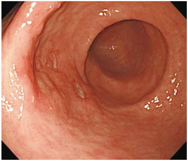
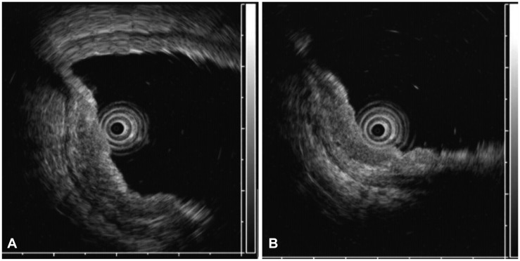
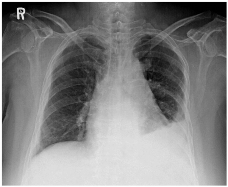
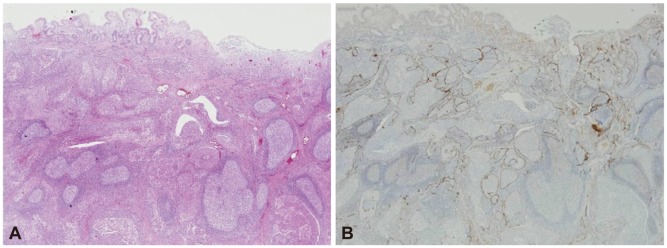
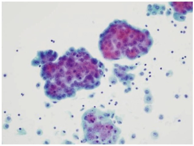
- The incidence of early gastric cancer (EGC) has increased to >50% in Korea owing to a higher detection rate caused by rapid advances in diagnostic instrumentation. EGC with distant metastasis has been rarely reported. Here, we report the case of a 76-year-old woman in whom general EGC was initially diagnosed by endoscopy and endoscopic ultrasonography. She subsequently underwent endoscopic submucosal dissection (ESD). Histological examination of the ESD specimen revealed that neoplastic cells were located predominantly in the submucosal layer and submucosal lymphatic channels. Metastatic cancer cells were also found in the pleural effusion. After conducting all analyses, including immunohistochemical staining, we concluded that the patient had primary EGC with pleural metastasis.
MeSH Terms
Figure
Reference
-
1. Ahn YO. Cancer in Korea: present features. Jpn J Clin Oncol. 2002; 32(Suppl):S32–S36. PMID: 11959875.
Article2. Gotoda T. Endoscopic resection of early gastric cancer. Gastric Cancer. 2007; 10:1–11. PMID: 17334711.
Article3. Hirasawa T, Gotoda T, Miyata S, et al. Incidence of lymph node metastasis and the feasibility of endoscopic resection for undifferentiated-type early gastric cancer. Gastric Cancer. 2009; 12:148–152. PMID: 19890694.
Article4. Adachi Y, Shiraishi N, Kitano S. Modern treatment of early gastric cancer: review of the Japanese experience. Dig Surg. 2002; 19:333–339. PMID: 12435900.
Article5. Ballantyne KC, Morris DL, Jones JA, Gregson RH, Hardcastle JD. Accuracy of identification of early gastric cancer. Br J Surg. 1987; 74:618–619. PMID: 3620874.
Article6. Sano T, Okuyama Y, Kobori O, Shimizu T, Morioka Y. Early gastric cancer. Endoscopic diagnosis of depth of invasion. Dig Dis Sci. 1990; 35:1340–1344. PMID: 2226095.7. Tsendsuren T, Jun SM, Mian XH. Usefulness of endoscopic ultrasonography in preoperative TNM staging of gastric cancer. World J Gastroenterol. 2006; 12:43–47. PMID: 16440415.
Article8. Nakazawa S. Recent advances in endoscopic ultrasonography. J Gastroenterol. 2000; 35:257–260. PMID: 10777153.
Article9. Kajitani T. The general rules for the gastric cancer study in surgery and pathology. Part I. Clinical classification. Jpn J Surg. 1981; 11:127–139. PMID: 7300058.10. Ohta H, Noguchi Y, Takagi K, Nishi M, Kajitani T, Kato Y. Early gastric carcinoma with special reference to macroscopic classification. Cancer. 1987; 60:1099–1106. PMID: 3607727.
Article11. Matsumoto Y, Yanai H, Tokiyama H, Nishiaki M, Higaki S, Okita K. Endoscopic ultrasonography for diagnosis of submucosal invasion in early gastric cancer. J Gastroenterol. 2000; 35:326–331. PMID: 10832666.
Article12. Shiomi M, Kamisako T, Yutani I, et al. Two cases of histopathologically advanced (stage IV) early gastric cancers. Tumori. 2001; 87:191–195. PMID: 11504376.
Article13. Sahn SA. Pleural diseases related to metastatic malignancies. Eur Respir J. 1997; 10:1907–1913. PMID: 9272937.
Article14. Lee MJ, Lee HS, Kim WH, Choi Y, Yang M. Expression of mucins and cytokeratins in primary carcinomas of the digestive system. Mod Pathol. 2003; 16:403–410. PMID: 12748245.
Article
- Full Text Links
- Actions
-
Cited
- CITED
-
- Close
- Share
- Similar articles
-
- Adenocarcinoma of Lung Cancer with Solitary Metastasis to the Stomach
- Early Gastric Cancer with Neurofibroma Mimicking a Metastatic Node: A Case Report
- Multiple Early Gastric Cancer
- Early Gastric Mucosal Cancer Associated with Synchronous Liver Metastasis
- Significance of Lymph Node Metastasis in Early Gastric Cancer

