A Case of Langerhans Cell Histiocytosis Manifested as a Suprasellar Mass
- Affiliations
-
- 1Center for Pediatric Cancer, National Cancer Center, Goyang, Korea. hjpark@ncc.re.kr
- 2Neuro-Oncology Clinic, National Cancer Center, Goyang, Korea.
- 3Department of Diagnostic Radiology, National Cancer Center, Goyang, Korea.
- 4Department of Pathology, National Cancer Center, Goyang, Korea.
- KMID: 2165231
- DOI: http://doi.org/10.14791/btrt.2016.4.1.26
Abstract
- Langerhans cell histiocytosis (LCH) has diverse clinical manifestations, including intracranial mass lesions. We report a case of LCH that manifested as a suprasellar mass, and initially misdiagnosed as a germ cell tumor. A 29-year-old woman presented with polyuria, polydipsia and amenorrhea. Laboratory findings revealed hypopituitarism with central diabetes insipidus, and a suprasellar mass and a pineal mass were observed on magnetic resonance imaging. Under the clinical impression of a germ cell tumor, the patient was treated with germ cell tumor chemotherapy (cisplatin and etoposide) and radiation therapy without biopsy. After initial shrinkage of the lesions, further growth of the tumor was observed and a biopsy was performed. The histopathology revealed LCH. After chemotherapy according to the LCH III protocol, the tumor disappeared. She is on regular follow up for 5 years without relapse. The present findings indicate that LCH should be included in the differential diagnosis of a suprasellar mass, even in adults, especially when it manifests with diabetes insipidus. This case also underscores the importance of a histopathologic diagnosis in patients with suprasellar tumors before the initiation of a specific therapy, even if the clinical findings are highly suggestive of a specific diagnosis.
Keyword
MeSH Terms
-
Adult
Amenorrhea
Biopsy
Central Nervous System Neoplasms
Diabetes Insipidus
Diabetes Insipidus, Neurogenic
Diagnosis
Diagnosis, Differential
Drug Therapy
Female
Follow-Up Studies
Germinoma
Histiocytosis, Langerhans-Cell*
Humans
Hypopituitarism
Magnetic Resonance Imaging
Neoplasms, Germ Cell and Embryonal
Polydipsia
Polyuria
Recurrence
Sella Turcica
Figure
-
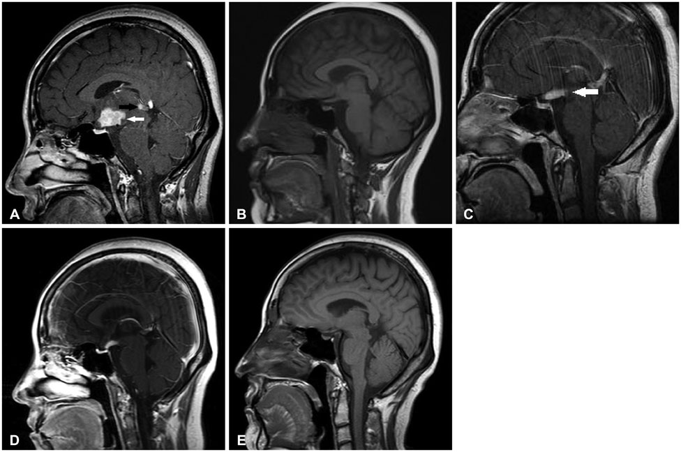
Fig. 1 Sagittal, T1-weighted images. A: Initial MRI on June 2007. Well enhanced mass with a diameter of 2.3 cm is observed around the pituitary stalk (white arrow). Another 1 cm-sized rim-enhancing lesion is observed in the pineal gland (black arrow). B: MRI on September 2007. After 3 cycles of germ cell tumor chemotherapy, mass size decreased markedly, showing only linear enhancement. C: MRI on September 2008. Increased size of enhancing mass in pituitary stalk and hypophysis areas observed (white arrow). D: MRI on December 2008. After Langerhans cell histiocytosis initial chemotherapy. Significant reduction in tumor size is observed. E: MRI on May 2015. Subtle residual enhancement in 3rd ventricle floor is observed. No other abnormal enhancing lesion is observed. Pituitary gland and stalk shows atrophy.

Fig. 2 Photograph of the surgical specimen. A: Many lymphohistiocytic infiltrations are noted in the low power view (HE staining, ×100). B: Scattered Langerhans cells are seen, with eosinophils and lymphocytes (HE staining, ×400). C and D: Immunohistochemical staining shows strong CD1 positivity (C) and weak S-100 positivity (D). HE, hematoxylin and eosin.
Cited by 1 articles
-
Pineal and Suprasellar Germinoma Cooccurence with Vertebra Plana: A Case Report
Farrokh Seilanian Toosi, Behzad Aminzadeh, Mohammad Faraji Rad, Sirous Nekooei, Mahsa Nahidi, Ehsan Keykhosravi
Brain Tumor Res Treat. 2018;6(2):73-77. doi: 10.14791/btrt.2018.6.e9.
Reference
-
1. Hershey BL. Suprasellar masses: diagnosis and differential diagnosis. Semin Ultrasound CT MR. 1993; 14:215–231.
Article2. Favara BE, Jaffe R. The histopathology of Langerhans cell histiocytosis. Br J Cancer Suppl. 1994; 23:S17–S23.3. Grois N, Pötschger U, Prosch H, et al. Risk factors for diabetes insipidus in langerhans cell histiocytosis. Pediatr Blood Cancer. 2006; 46:228–233.
Article4. Baumgartner I, von Hochstetter A, Baumert B, Luetolf U, Follath F. Langerhans'-cell histiocytosis in adults. Med Pediatr Oncol. 1997; 28:9–14.
Article5. Vassallo R, Ryu JH, Colby TV, Hartman T, Limper AH. Pulmonary Langerhans'-cell histiocytosis. N Engl J Med. 2000; 342:1969–1978.
Article6. Prayer D, Grois N, Prosch H, Gadner H, Barkovich AJ. MR imaging presentation of intracranial disease associated with Langerhans cell histiocytosis. AJNR Am J Neuroradiol. 2004; 25:880–891.7. Echevarría ME, Fangusaro J, Goldman S. Pediatric central nervous system germ cell tumors: a review. Oncologist. 2008; 13:690–699.
Article8. Ono N, Isobe I, Uki J, Kurihara H, Shimizu T, Kohno K. Recurrence of primary intracranial germinomas after complete response with radiotherapy: recurrence patterns and therapy. Neurosurgery. 1994; 35:615–620.9. Sawamura Y, de Tribolet N, Ishii N, Abe H. Management of primary intracranial germinomas: diagnostic surgery or radical resection? J Neurosurg. 1997; 87:262–266.
Article10. Tien RD, Newton TH, McDermott MW, Dillon WP, Kucharczyk J. Thickened pituitary stalk on MR images in patients with diabetes insipidus and Langerhans cell histiocytosis. AJNR Am J Neuroradiol. 1990; 11:703–708.11. Prosch H, Grois N, Bökkerink J, et al. Central diabetes insipidus: is it Langerhans cell histiocytosis of the pituitary stalk? A diagnostic pitfall. Pediatr Blood Cancer. 2006; 46:363–366.
Article12. Buckner JC, Peethambaram PP, Smithson WA, et al. Phase II trial of primary chemotherapy followed by reduced-dose radiation for CNS germ cell tumors. J Clin Oncol. 1999; 17:933–940.
Article13. Yu LC, Shenoy S, Ward K, Warrier RP. Successful treatment of multisystem Langerhans cell histiocytosis (histiocytosis X) with etoposide. Am J Pediatr Hematol Oncol. 1994; 16:275–277.
Article
- Full Text Links
-
- Actions
-
Cited
- CITED
-
- Close
- Share
-
- Similar articles
-
- A Case of Orbital Langerhans' cell histiocytosis
- Two Cases of Langerhans' Cell Histiocytosis: Report of Two Cases
- Spontaneous Pneumothorax due to Pulmonary Invasion in Multisystemic Langerhans Cell Histiocytosis: A case report
- A Case of Langerhans Cell Histiocytosis Mimicking Periorbital Cellulitis
- Pulmonary Langerhans Cell Histiocytosis Accompanied by Active Pulmonary Tuberculosis
