Dynamic Enhanced Computed Tomographic Findings of a Perirenal Capillary Hemangioma
- Affiliations
-
- 1Department of Radiology, Kyung Hee University Hospital at Gangdong, Seoul, Korea. rad2000@hanmail.net
- 2Department of Pathology, Kyung Hee University Hospital at Gangdong, Seoul, Korea.
- KMID: 2164828
- DOI: http://doi.org/10.3348/jksr.2016.74.5.322
Abstract
- Hemangiomas are benign mesenchymal neoplasms that rarely occur in the kidney and perirenal space. Perirenal hemangiomas can mimic the appearance of exophytic renal cell carcinoma or various retroperitoneal tumors. We report a case of perirenal hemangioma detected by dynamic enhanced computed tomography in a 43-year-old female.
MeSH Terms
Figure
-
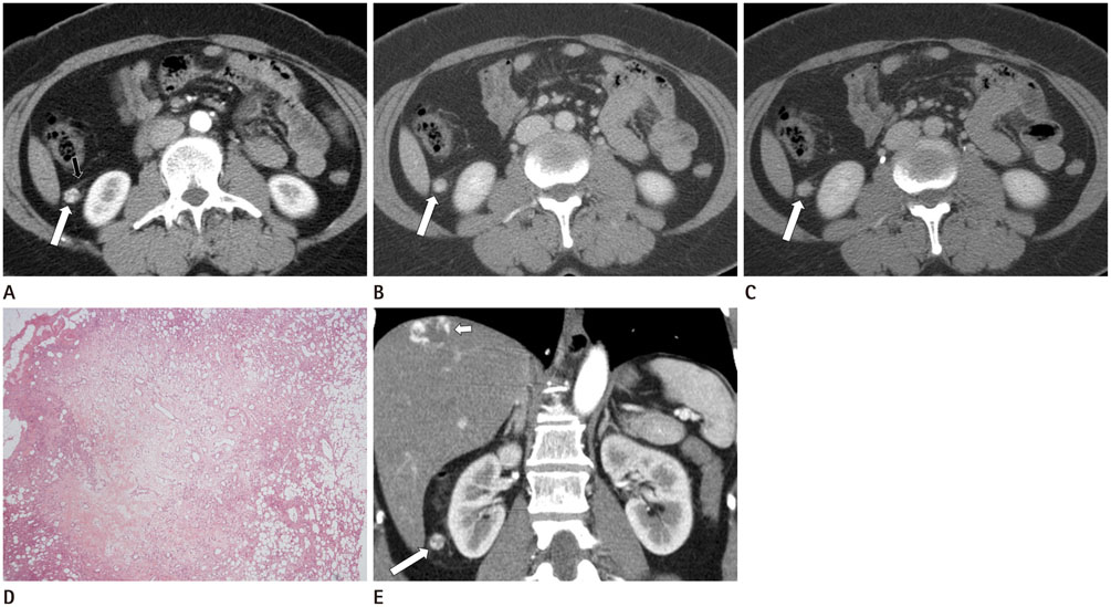
Fig. 1 Fig. 1. A 43-year-old woman with a capillary hemangioma in the perirenal space. A. Contrast-enhanced arterial phase CT showing a small, peripheral-enhancing mass (arrow) in the right perirenal space. There is a mild thickening of the anterior perirenal fascia (small arrow) adjacent to the mass. B, C. Contrast-enhanced CT images of the venous phase (B) and delayed phase (C) revealing homogeneous enhancement of the perirenal mass (arrows). The mass does not adhere to the surface of the right kidney. It is accompanied by mild fat stranding. D. Photomicrograph of the perirenal mass showing the proliferation of small capillary vessels (hematoxylin and eosin staining, × 20). The mass is surrounded by perirenal fat tissue. E. Coronal reformatted image of enhanced arterial phase CT revealing a small mass (arrow) with peripheral globular enhancement (short arrow), similar to that seen in hepatic hemangioma, around the right kidney.
Reference
-
1. Okuno T, Ando M, Arisawa C, Okano T. A case of perirenal hemangioma mimicking renal cell carcinoma. Int J Urol. 1999; 6:104–106.2. Wang T, Palazzo JP, Mitchell D, Petersen RO. Renal capsular hemangioma. J Urol. 1993; 149:1122–1123.3. Higuchi R, Yamaguchi Y, Shoji T, Wakasugi S, Takahashi H, Fujita R. A mediastinal hemangioma, associated with perirenal hemangioma and congenital anomaly of the inferior vena cava. Intern Med. 2000; 39:1083–1087.4. Surabhi VR, Menias C, Prasad SR, Patel AH, Nagar A, Dalrymple NC. Neoplastic and non-neoplastic proliferative disorders of the perirenal space: cross-sectional imaging findings. Radiographics. 2008; 28:1005–1017.5. Prasad SR, Humphrey PA, Catena JR, Narra VR, Srigley JR, Cortez AD, et al. Common and uncommon histologic subtypes of renal cell carcinoma: imaging spectrum with pathologic correlation. Radiographics. 2006; 26:1795–1806. discussion 1806-18106. Neville A, Herts BR. CT characteristics of primary retroperitoneal neoplasms. Crit Rev Comput Tomogr. 2004; 45:247–270.7. Obara W, Sato K, Owari Y, Nozawa T, Isurugi K, Ohmori S, et al. Perinephric angiomyolipoma: a unique development pattern surrounding the kidney. Int J Urol. 2005; 12:305–307.8. Enomoto K, Nakamichi I, Hamada K, Inoue A, Higuchi I, Sekimoto M, et al. Unicentric and multicentric Castleman's disease. Br J Radiol. 2007; 80:e24–e26.9. Lee HS, Koh BH, Kim JW, Kim YS, Rhim HC, Cho OK, et al. Radiologic findings of renal hemangioma: report of three cases. Korean J Radiol. 2000; 1:60–63.10. Prasad SR, Humphrey PA, Menias CO, Middleton WD, Siegel MJ, Bae KT, et al. Neoplasms of the renal medulla: radiologic-pathologic correlation. Radiographics. 2005; 25:369–380.
- Full Text Links
-
- Actions
-
Cited
- CITED
-
- Close
- Share
-
- Similar articles
-
- A Suspicious Breast Lesion Detected by Dynamic Contrast-Enhanced MRI and Pathologically Confirmed as Capillary Hemangioma: a Case Report and Literature Review
- Recurrent Pulmonary Capillary Hemangioma: Dynamic Contrast-Enhanced CT and Histopathologic Findings
- Capillary Hemangioma of Testis
- Cutis Marmorata Telangiectatica Congenita: A Rare Clinical Manifestation of Capillary Hemangioma?
- Anastomosing Hemangioma Involving the Para-Aortic Region: A Case Report
