Recurrent Pulmonary Capillary Hemangioma: Dynamic Contrast-Enhanced CT and Histopathologic Findings
- Affiliations
-
- 1Department of Radiology and Center for Imaging Science, Samsung Medical Center, Sungkyunkwan University School of Medicine, Seoul 135-710, Korea. tskim.kim@samsung.com
- 2Department of Pathology, Samsung Medical Center, Sungkyunkwan University School of Medicine, Seoul 135-710, Korea.
- 3Division of Pulmonary and Critical Care Medicine, Department of Medicine, Sungkyunkwan University School of Medicine, Seoul 135-710, Korea.
- 4Department of Thoracic Surgery, Samsung Medical Center, Sungkyunkwan University School of Medicine, Seoul 135-710, Korea.
- 5Department of Radiology, Gachon University Gil Hospital, Incheon 405-760, Korea.
- KMID: 1372856
- DOI: http://doi.org/10.3348/kjr.2012.13.3.350
Abstract
- We report the dynamic contrast-enhanced CT and histopathologic findings of a rare case of recurrent pulmonary capillary hemangiomas. The findings consisted of peripheral nodular enhancement at the early arterial phase and a subsequent "central filling-in" enhancement pattern on the delayed scans, which was identical to the well-known enhancement pattern of hemangiomas of the liver. Although there was no evidence of histological malignancy, pulmonary capillary hemangiomas manifested as multiple nodular lesions and showed postoperative recurrence.
Keyword
MeSH Terms
Figure
-
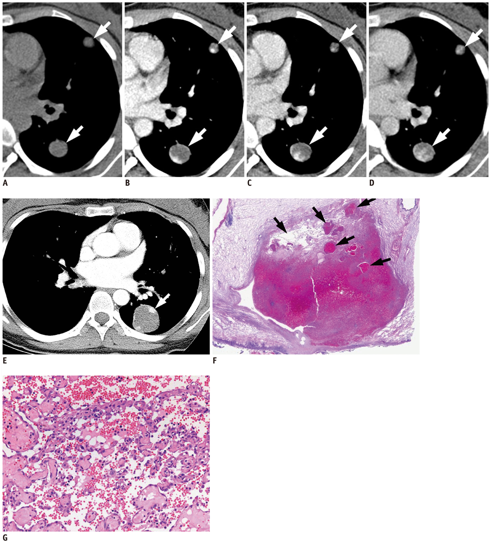
Fig. 1 Pulmonary capillary hemangiomas in 22-year-old female. Initial multi-phase dymamic CT scan obtained at outside hospital shows two pulmonary nodules in left lung. Dynamic postcontrast CT scans (B-D) show peripheral nodular enhancement in early phase CT (B) and "central filling-in" enhancement pattern of theses nodules in subsequent delayed images (C, D), which are identical to well-known enhancement pattern of hepatic hemangiomas (A: precontrast image, B: obtained 1 minute after contrast enhancement, C: 2 minutes, D: 4 minutes). Wedge resection of these left pulmonary nodules was performed at outside hospital, and review of pathologic specimen revealed pulmonary capillary hemangiomas. Initial multi-phase dynamic CT scan obtained at outside hospital shows two pulmonary nodules in left lung. E. Fifty months after tumor removal, chest CT scan obtained from our institution shows 30-mm, recurrent pulmonary nodule in left lower lobe. Another smaller nodule is also detected in right lower lobe (not shown here). Nodule in left lower lobe shows prominent "peripheral nodular enhancement" pattern at this post-contrast CT image obtained 40 seconds after contrast administration, which is identical to previous CT finding. F. Scanning photograph of histopathologic specimen obtained from left lower lobectomy shows well-demarcated hemorrhagic nodule containing multiple dilated vascular spaces (arrows), many of which are compactly packed with red blood cells (H and E, ×1). G. Low-power photomicrograph shows that nodule is composed of anastomosing capillary vascular channels filled with red blood cells. These capillary lumina are divided by narrow trabeculae of hyaline stroma (H and E, ×40). These findings are compatible with capillary hemangiomas.
Reference
-
1. Bowyer JJ, Sheppard M. Capillary haemangioma presenting as a lung pseudocyst. Arch Dis Child. 1990. 65:1162–1164.2. Galliani CA, Beatty JF, Grosfeld JL. Cavernous hemangioma of the lung in an infant. Pediatr Pathol. 1992. 12:105–111.3. Abrahams NA, Colby TV, Pearl RH, Chipps BE, Juris AL, Leslie KO. Pulmonary hemangiomas of infancy and childhood: report of two cases and review of the literature. Pediatr Dev Pathol. 2002. 5:283–292.4. Fugo K, Matsuno Y, Okamoto K, Kusumoto M, Maeshima A, Kaji M, et al. Solitary capillary hemangioma of the lung: report of 2 resected cases detected by high-resolution CT. Am J Surg Pathol. 2006. 30:750–753.5. Capizzani TR, Patel H, Hines MH, Mott RT, Petty JK. A unique case of a giant congenital pulmonary hemangioma in a newborn. J Pediatr Surg. 2008. 43:574–578.6. Dinehart SM, Kincannon J, Geronemus R. Hemangiomas: evaluation and treatment. Dermatol Surg. 2001. 27:475–485.7. Sienko A. Danis S, Zander , Farver CF, editors. Pulmonary processes of indeterminate malignant potential. Pulmonary pathology. 2008. Philadelphia, USA: Churchill Livingstone;636–648.8. Flieder DB. Zander Danis S., Farver CF, editors. Benign Neoplasms of the Lungs. Pulmonary pathology. 2008. Philadelphia, USA: Churchill Livingstone;669–672.9. Katzenstein AL, Gmelich JT, Carrington CB. Sclerosing hemangioma of the lung: a clinicopathologic study of 51 cases. Am J Surg Pathol. 1980. 4:343–356.10. Chung MJ, Lee KS, Han J, Sung YM, Chong S, Kwon OJ. Pulmonary sclerosing hemangioma presenting as solitary pulmonary nodule: dynamic CT findings and histopathologic comparisons. AJR Am J Roentgenol. 2006. 187:430–437.
- Full Text Links
-
- Actions
-
Cited
- CITED
-
- Close
- Share
-
- Similar articles
-
- A Suspicious Breast Lesion Detected by Dynamic Contrast-Enhanced MRI and Pathologically Confirmed as Capillary Hemangioma: a Case Report and Literature Review
- Hepatic Cavernous Hemangioma in Cirrhotic Liver: Imaging Findings
- Dynamic Enhanced Computed Tomographic Findings of a Perirenal Capillary Hemangioma
- Primary Lung Cancer: Utility of Contrast-enhanced Dynamic CT in Diagnosis with Histopathologic Correlation
- Dynamic CT Features of a Hemangioma Originating from the Parietal Pleura: A Case Report
