J Korean Soc Radiol.
2016 May;74(5):291-298. 10.3348/jksr.2016.74.5.291.
Coronary Stent on Coronary CT Angiography: Assessment with Model-Based Iterative Reconstruction Technique
- Affiliations
-
- 1Department of Radiology, Seoul National University Bundang Hospital, Seongnam, Korea. drsic@hanmail.net
- KMID: 2164824
- DOI: http://doi.org/10.3348/jksr.2016.74.5.291
Abstract
- PURPOSE
To assess the performance of model-based iterative reconstruction (MBIR) technique for evaluation of coronary artery stents on coronary CT angiography (CCTA).
MATERIALS AND METHODS
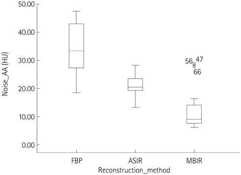
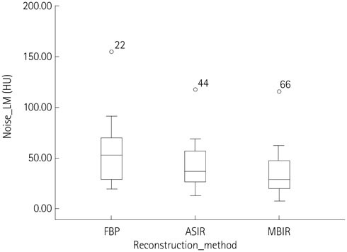
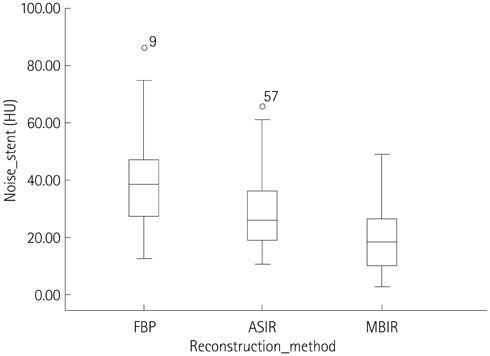
Twenty-two patients with coronary stent implantation who underwent CCTA were retrospectively enrolled for comparison of image quality between filtered back projection (FBP), adaptive statistical iterative reconstruction (ASIR) and MBIR. In each data set, image noise was measured as the standard deviation of the measured attenuation units within circular regions of interest in the ascending aorta (AA) and left main coronary artery (LM). To objectively assess the noise and blooming artifacts in coronary stent, we additionally measured the standard deviation of the measured attenuation and intra-luminal stent diameters of total 35 stents with dedicated software.
RESULTS
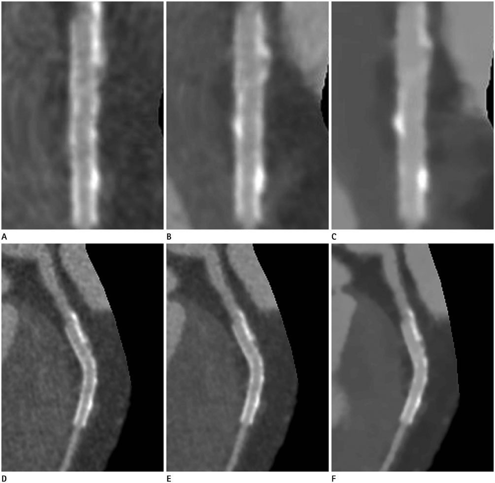
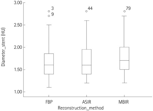
All image noise measured in the AA (all p < 0.001), LM (p < 0.001, p = 0.001) and coronary stent (all p < 0.001) were significantly lower with MBIR in comparison to those with FBP or ASIR. Intraluminal stent diameter was significantly higher with MBIR, as compared with ASIR or FBP (p < 0.001, p = 0.001).
CONCLUSION
MBIR can reduce image noise and blooming artifact from the stent, leading to better in-stent assessment in patients with coronary artery stent.
MeSH Terms
Figure
Reference
-
1. Ebersberger U, Tricarico F, Schoepf UJ, Blanke P, Spears JR, Rowe GW, et al. CT evaluation of coronary artery stents with iterative image reconstruction: improvements in image quality and potential for radiation dose reduction. Eur Radiol. 2013; 23:125–132.2. Kim YJ, Yong HS, Kim SM, Kim JA, Yang DH, Hong YJ. Guideline for Appropriate Use of Cardiac CT in Heart Disease. J Korean Soc Radiol. 2014; 70:93–109.3. Taylor AJ, Cerqueira M, Hodgson JM, Mark D, Min J, O'Gara P, et al. ACCF/SCCT/ACR/AHA/ASE/ASNC/NASCI/SCAI/SCMR 2010 appropriate use criteria for cardiac computed tomography. A report of the American College of Cardiology Foundation Appropriate Use Criteria Task Force, the Society of Cardiovascular Computed Tomography, the American College of Radiology, the American Heart Association, the American Society of Echocardiography, the American Society of Nuclear Cardiology, the North American Society for Cardiovascular Imaging, the Society for Cardiovascular Angiography and Interventions, and the Society for Cardiovascular Magnetic Resonance. J Am Coll Cardiol. 2010; 56:1864–1894.4. Andreini D, Pontone G, Bartorelli AL, Mushtaq S, Trabattoni D, Bertella E, et al. High diagnostic accuracy of prospective ECG-gating 64-slice computed tomography coronary angiography for the detection of in-stent restenosis: in-stent restenosis assessment by low-dose MDCT. Eur Radiol. 2011; 21:1430–1438.5. Kerl JM, Schoepf UJ, Vogl TJ, Ackermann H, Vogt S, Costello P, et al. In vitro evaluation of metallic coronary artery stents with 64-MDCT using an ECG-gated cardiac phantom: relationship between in-stent visualization, stent type, and heart rate. AJR Am J Roentgenol. 2010; 194:W256–W262.6. Sheth T, Dodd JD, Hoffmann U, Abbara S, Finn A, Gold HK, et al. Coronary stent assessability by 64 slice multi-detector computed tomography. Catheter Cardiovasc Interv. 2007; 69:933–938.7. Zhang X, Yang L, Ju H, Zhang F, Wu J, He B, et al. Prevalence and prognosis of coronary stent gap detected by multi-detector CT: a follow-up study. Eur Radiol. 2012; 22:1896–1903.8. Sun Z, Almutairi AM. Diagnostic accuracy of 64 multislice CT angiography in the assessment of coronary in-stent restenosis: a meta-analysis. Eur J Radiol. 2010; 73:266–273.9. Kumbhani DJ, Ingelmo CP, Schoenhagen P, Curtin RJ, Flamm SD, Desai MY. Meta-analysis of diagnostic efficacy of 64-slice computed tomography in the evaluation of coronary in-stent restenosis. Am J Cardiol. 2009; 103:1675–1681.10. Gosling O, Loader R, Venables P, Roobottom C, Rowles N, Bellenger N, et al. A comparison of radiation doses between state-of-the-art multislice CT coronary angiography with iterative reconstruction, multislice CT coronary angiography with standard filtered back-projection and invasive diagnostic coronary angiography. Heart. 2010; 96:922–926.11. Prakash P, Kalra MK, Ackman JB, Digumarthy SR, Hsieh J, Do S, et al. Diffuse lung disease: CT of the chest with adaptive statistical iterative reconstruction technique. Radiology. 2010; 256:261–269.12. Sagara Y, Hara AK, Pavlicek W, Silva AC, Paden RG, Wu Q. Abdominal CT: comparison of low-dose CT with adaptive statistical iterative reconstruction and routine-dose CT with filtered back projection in 53 patients. AJR Am J Roentgenol. 2010; 195:713–719.13. Renker M, Nance JW Jr, Schoepf UJ, O'Brien TX, Zwerner PL, Meyer M, et al. Evaluation of heavily calcified vessels with coronary CT angiography: comparison of iterative and filtered back projection image reconstruction. Radiology. 2011; 260:390–399.14. Funama Y, Oda S, Utsunomiya D, Taguchi K, Shimonobo T, Yamashita Y, et al. Coronary artery stent evaluation by combining iterative reconstruction and high-resolution kernel at coronary CT angiography. Acad Radiol. 2012; 19:1324–1331.15. Scheffel H, Stolzmann P, Schlett CL, Engel LC, Major GP, Károlyi M, et al. Coronary artery plaques: cardiac CT with model-based and adaptive-statistical iterative reconstruction technique. Eur J Radiol. 2012; 81:e363–e369.16. Katsura M, Sato J, Akahane M, Matsuda I, Ishida M, Yasaka K, et al. Comparison of pure and hybrid iterative reconstruction techniques with conventional filtered back projection: image quality assessment in the cervicothoracic region. Eur J Radiol. 2013; 82:356–360.17. Chang W, Lee JM, Lee K, Yoon JH, Yu MH, Han JK, et al. Assessment of a model-based, iterative reconstruction algorithm (MBIR) regarding image quality and dose reduction in liver computed tomography. Invest Radiol. 2013; 48:598–606.18. Kim H, Park CM, Song YS, Lee SM, Goo JM. Influence of radiation dose and iterative reconstruction algorithms for measurement accuracy and reproducibility of pulmonary nodule volumetry: a phantom study. Eur J Radiol. 2014; 83:848–857.19. Lee MS, Chun EJ, Kim KJ, Kim JA, Vembar M, Choi SI. Reproducibility in the assessment of noncalcified coronary plaque with 256-slice multi-detector CT and automated plaque analysis software. Int J Cardiovasc Imaging. 2010; 26:Suppl 2. 237–244.20. Chun EJ, Lee W, Choi YH, Koo BK, Choi SI, Jae HJ, et al. Effects of nitroglycerin on the diagnostic accuracy of electrocardiogram-gated coronary computed tomography angiography. J Comput Assist Tomogr. 2008; 32:86–92.21. Oda S, Utsunomiya D, Funama Y, Katahira K, Honda K, Tokuyasu S, et al. A knowledge-based iterative model reconstruction algorithm: can super-low-dose cardiac CT be applicable in clinical settings? Acad Radiol. 2014; 21:104–110.22. Shuman WP, Green DE, Busey JM, Kolokythas O, Mitsumori LM, Koprowicz KM, et al. Model-based iterative reconstruction versus adaptive statistical iterative reconstruction and filtered back projection in liver 64-MDCT: focal lesion detection, lesion conspicuity, and image noise. AJR Am J Roentgenol. 2013; 200:1071–1076.
- Full Text Links
- Actions
-
Cited
- CITED
-
- Close
- Share
- Similar articles
-
- Erratum: Coronary Stent on Coronary CT Angiography: Assessment with Model-Based Iterative Reconstruction Technique
- Coronary CT AngiographyBased Assessment of Coronary in-Stent Restenosis: A Journey through Past and Present Trends
- Assessment of Coronary Stenosis Using Coronary CT Angiography in Patients with High Calcium Scores: Current Limitations and Future Perspectives
- Effects of Iterative Reconstruction Algorithm, Automatic Exposure Control on Image Quality, and Radiation Dose: Phantom Experiments with Coronary CT Angiography Protocols
- Coronary CT Angiography

