Breast Magnetic Resonance Imaging-Guided Biopsy
- Affiliations
-
- 1Department of Radiology, Seoul National University Bundang Hospital, Seongnam, Korea. kimsmlms@daum.net
- 2Department of Radiology, Seoul National University Hospital, Seoul, Korea.
- 3Department of Radiology, Asan Medical Center, University of Ulsan College of Medicine, Seoul, Korea.
- KMID: 2164811
- DOI: http://doi.org/10.3348/jksr.2016.74.6.351
Abstract
- Despite the high sensitivity of breast magnetic resonance imaging (MRI), pathologic confirmation by biopsy is essential because of limited specificity. MRI-guided biopsy is required in patients with lesions only seen on MRI. We review preprocedural considerations and the technique of MRI-guided biopsy, challenging situations and trouble-shooting, and correlation of radiologic and pathologic findings.
MeSH Terms
Figure
-

Fig. 1 MRI-guided biopsy grid system.

Fig. 2 MRI-guided biopsy kit contains introducer stylet, obturator, introducer sheath, and needle guide.

Fig. 3 MRI-guided VAB procedure. After localizing image (A), precontrast images with fiducial marker (arrow) (B) are obtained. Sagittal and axial postcontrast images (C, D) are obtained to identify target location (arrow). VAB = vacuum assisted biopsy After location of introducer sheath and obturator, sagittal and axial images (E, F) are obtained to confirm the position before lesion sampling. The target lesion (arrowhead) and obturator (arrow) are well demonstrated in these images. After tissue sampling, additional sagittal and axial images (G, H) are obtained to confirm adequate biopsy location and marker (arrow) placement. The target lesion was confirmed as ductal carcinoma in situ. VAB = vacuum assisted biopsy

Fig. 4 Example of worksheet.
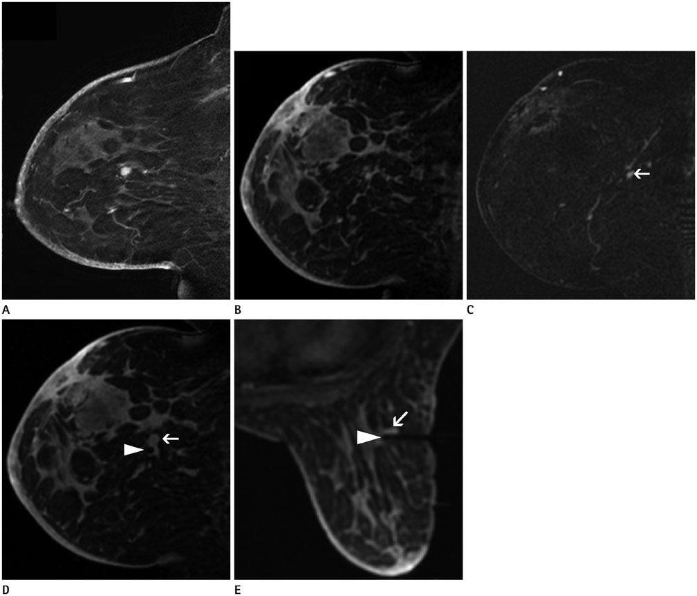
Fig. 5 A 35-year-old woman with ipsilateral breast cancer surgery 9 months previously. Postoperative MRI shows a new, round, fast and washout-enhancing mass (A). The patient underwent MRI-guided biopsy. The target lesion is not identified on the postcontrast sagittal image (B). On the subtraction image (C), a subtle enhancing mass is well-delineated (arrow). Repeat MRI (D, E) shows introducer sheath with obturator (arrowhead) in correct position, with the tip at the target lesion (arrow). The enhancing mass was pathologically confirmed as reactive hyperplasia in intramammary lymph node.

Fig. 6 Lateral approach for posteromedial located target.
Reference
-
1. Kuhl C, Weigel S, Schrading S, Arand B, Bieling H, König R, et al. Prospective multicenter cohort study to refine management recommendations for women at elevated familial risk of breast cancer: the EVA trial. J Clin Oncol. 2010; 28:1450–1457.2. Berg WA, Zhang Z, Lehrer D, Jong RA, Pisano ED, Barr RG, et al. Detection of breast cancer with addition of annual screening ultrasound or a single screening MRI to mammography in women with elevated breast cancer risk. JAMA. 2012; 307:1394–1404.3. Morris EA, Schwartz LH, Dershaw DD, van Zee KJ, Abramson AF, Liberman L. MR imaging of the breast in patients with occult primary breast carcinoma. Radiology. 1997; 205:437–440.4. Boetes C, Mus RD, Holland R, Barentsz JO, Strijk SP, Wobbes T, et al. Breast tumors: comparative accuracy of MR imaging relative to mammography and US for demonstrating extent. Radiology. 1995; 197:743–747.5. Gavenonis SC, Roth SO. Role of magnetic resonance imaging in evaluating the extent of disease. Magn Reson Imaging Clin N Am. 2010; 18:199–206. vii–viii.6. Mann RM. The effectiveness of MR imaging in the assessment of invasive lobular carcinoma of the breast. Magn Reson Imaging Clin N Am. 2010; 18:259–276. ix7. Pediconi F, Catalano C, Roselli A, Padula S, Altomari F, Moriconi E, et al. Contrast-enhanced MR mammography for evaluation of the contralateral breast in patients with diagnosed unilateral breast cancer or high-risk lesions. Radiology. 2007; 243:670–680.8. Wang SY, Long JB, Killelea BK, Evans SB, Roberts KB, Silber A, et al. Preoperative breast magnetic resonance imaging and contralateral breast cancer occurrence among older women with breast cancer. J Clin Oncol. 2016; 34:321–328.9. Iacconi C, Galman L, Zheng J, Sacchini V, Sutton EJ, Dershaw D, et al. Multicentric cancer detected at breast MR imaging and not at mammography: important or not? Radiology. 2016; 279:378–384.10. Peters NH, Borel Rinkes IH, Zuithoff NP, Mali WP, Moons KG, Peeters PH. Meta-analysis of MR imaging in the diagnosis of breast lesions. Radiology. 2008; 246:116–124.11. American College of Radiology. ACR practice parameter for the performance of contrast-enhanced magnetic resonance imaging (MRI) of the breast. 2014. Accessed May 4, 2016. Available at: http://www.acr.org/~/media/ACR/Documents/PGTS/guidelines/MRI_Breast.pdf.12. Mann RM, Kuhl CK, Kinkel K, Boetes C. Breast MRI: guidelines from the European Society of Breast Imaging. Eur Radiol. 2008; 18:1307–1318.13. Jung HN, Han BK, Ko EY, Shin JH. Initial experience with magnetic resonance-guided vacuum-assisted biopsy in Korean women with breast cancer. J Breast Cancer. 2014; 17:270–278.14. Choi HY, Kim SM, Jang M, Yun BL, Kim SW, Kang E, et al. MRI-guided intervention for breast lesions using the freehand technique in a 3.0-T closed-bore MRI scanner: feasibility and initial results. Korean J Radiol. 2013; 14:171–178.15. Abe H, Schmidt RA, Shah RN, Shimauchi A, Kulkarni K, Sennett CA, et al. MR-directed ("Second-Look") ultrasound examination for breast lesions detected initially on MRI: MR and sonographic findings. AJR Am J Roentgenol. 2010; 194:370–377.16. LaTrenta LR, Menell JH, Morris EA, Abramson AF, Dershaw DD, Liberman L. Breast lesions detected with MR imaging: utility and histopathologic importance of identification with US. Radiology. 2003; 227:856–861.17. Meissnitzer M, Dershaw DD, Lee CH, Morris EA. Targeted ultrasound of the breast in women with abnormal MRI findings for whom biopsy has been recommended. AJR Am J Roentgenol. 2009; 193:1025–1029.18. Lee SH, Kim SM, Jang M, Yun BL, Kang E, Kim SW, et al. Role of second-look ultrasound examinations for MR-detected lesions in patients with breast cancer. Ultraschall Med. 2015; 36:140–148.19. Perlet C, Heywang-Kobrunner SH, Heinig A, Sittek H, Casselman J, Anderson I, et al. Magnetic resonance-guided, vacuum-assisted breast biopsy: results from a European multicenter study of 538 lesions. Cancer. 2006; 106:982–990.20. Heywang-Köbrunner SH, Sinnatamby R, Lebeau A, Lebrecht A, Britton PD, Schreer I. Consensus Group. Interdisciplinary consensus on the uses and technique of MR-guided vacuum-assisted breast biopsy (VAB): results of a European consensus meeting. Eur J Radiol. 2009; 72:289–294.21. Hefler L, Casselman J, Amaya B, Heinig A, Alberich T, Koelbl H, et al. Follow-up of breast lesions detected by MRI not biopsied due to absent enhancement of contrast medium. Eur Radiol. 2003; 13:344–346.22. Brennan SB, Sung JS, Dershaw DD, Liberman L, Morris EA. Cancellation of MR imaging-guided breast biopsy due to lesion nonvisualization: frequency and follow-up. Radiology. 2011; 261:92–99.23. Perlet C, Heinig A, Prat X, Casselman J, Baath L, Sittek H, et al. Multicenter study for the evaluation of a dedicated biopsy device for MR-guided vacuum biopsy of the breast. Eur Radiol. 2002; 12:1463–1470.24. Liberman L, Morris EA, Dershaw DD, Thornton CM, Van Zee KJ, Tan LK. Fast MRI-guided vacuum-assisted breast biopsy: initial experience. AJR Am J Roentgenol. 2003; 181:1283–1293.25. Price ER. Magnetic resonance imaging-guided biopsy of the breast: fundamentals and finer points. Magn Reson Imaging Clin N Am. 2013; 21:571–581.26. Liberman L. Percutaneous image-guided core breast biopsy. Radiol Clin North Am. 2002; 40:483–500. vi27. Bahrs SD, Hattermann V, Preibsch H, Hahn M, Staebler A, Claussen CD, et al. MR imaging-guided vacuum-assisted breast biopsy: reduction of false-negative biopsies by short-term control MRI 24-48 h after biopsy. Clin Radiol. 2014; 69:695–702.28. Crystal P, Sadaf A, Bukhanov K, McCready D, O'alley F, Helbich TH. High-risk lesions diagnosed at MRI-guided vacuum-assisted breast biopsy: can underestimation be predicted? Eur Radiol. 2011; 21:582–589.29. Gebauer B, Bostanjoglo M, Moesta KT, Schneider W, Schlag PM, Felix R. Magnetic resonance-guided biopsy of suspicious breast lesions with a handheld vacuum biopsy device. Acta Radiol. 2006; 47:907–913.30. Han BK, Schnall MD, Orel SG, Rosen M. Outcome of MRIguided breast biopsy. AJR Am J Roentgenol. 2008; 191:1798–1804.31. Hauth EA, Jaeger HJ, Lubnau J, Maderwald S, Otterbach F, Kimmig R, et al. MR-guided vacuum-assisted breast biopsy with a handheld biopsy system: clinical experience and results in postinterventional MR mammography after 24 h. Eur Radiol. 2008; 18:168–176.32. Kuhl CK, Morakkabati N, Leutner CC, Schmiedel A, Wardelmann E, Schild HH. MR imaging--guided large-core (14-gauge) needle biopsy of small lesions visible at breast MR imaging alone. Radiology. 2001; 220:31–39.33. Liberman L, Bracero N, Morris E, Thornton C, Dershaw DD. MRI-guided 9-gauge vacuum-assisted breast biopsy: initial clinical experience. AJR Am J Roentgenol. 2005; 185:183–193.34. Malhaire C, El Khoury C, Thibault F, Athanasiou A, Petrow P, Ollivier L, et al. Vacuum-assisted biopsies under MR guidance: results of 72 procedures. Eur Radiol. 2010; 20:1554–1562.35. Meeuwis C, Veltman J, van Hall HN, Mus RD, Boetes C, Barentsz JO, et al. MR-guided breast biopsy at 3T: diagnostic yield of large core needle biopsy compared with vacuum-assisted biopsy. Eur Radiol. 2012; 22:341–349.36. Noroozian M, Gombos EC, Chikarmane S, Georgian-Smith D, Raza S, Denison CM, et al. Factors that impact the duration of MRI-guided core needle biopsy. AJR Am J Roentgenol. 2010; 194:W150–W157.37. Jackman RJ, Marzoni FA Jr. Stereotactic histologic biopsy with patients prone: technical feasibility in 98% of mammographically detected lesions. AJR Am J Roentgenol. 2003; 180:785–794.38. Orel SG, Rosen M, Mies C, Schnall MD. MR imaging-guided 9-gauge vacuum-assisted core-needle breast biopsy: initial experience. Radiology. 2006; 238:54–61.39. Rauch GM, Dogan BE, Smith TB, Liu P, Yang WT. Outcome analysis of 9-gauge MRI-guided vacuum-assisted core needle breast biopsies. AJR Am J Roentgenol. 2012; 198:292–299.40. Heller SL, Elias K, Gupta A, Greenwood HI, Mercado CL, Moy L. Outcome of high-risk lesions at MRI-guided 9-gauge vacuum-assisted breast biopsy. AJR Am J Roentgenol. 2014; 202:237–245.41. Li J, Dershaw DD, Lee CH, Kaplan J, Morris EA. MRI followup after concordant, histologically benign diagnosis of breast lesions sampled by MRI-guided biopsy. AJR Am J Roentgenol. 2009; 193:850–855.42. Liberman L, Holland AE, Marjan D, Murray MP, Bartella L, Morris EA, et al. Underestimation of atypical ductal hyperplasia at MRI-guided 9-gauge vacuum-assisted breast biopsy. AJR Am J Roentgenol. 2007; 188:684–690.43. Strigel RM, Eby PR, Demartini WB, Gutierrez RL, Allison KH, Peacock S, et al. Frequency, upgrade rates, and characteristics of high-risk lesions initially identified with breast MRI. AJR Am J Roentgenol. 2010; 195:792–798.44. Lee JM, Kaplan JB, Murray MP, Mazur-Grbec M, Tadic T, Stimac D, et al. Underestimation of DCIS at MRI-guided vacuum-assisted breast biopsy. AJR Am J Roentgenol. 2007; 189:468–474.45. Eby PR, Ochsner JE, DeMartini WB, Allison KH, Peacock S, Lehman CD. Frequency and upgrade rates of atypical ductal hyperplasia diagnosed at stereotactic vacuum-assisted breast biopsy: 9-versus 11-gauge. AJR Am J Roentgenol. 2009; 192:229–234.46. Jackman RJ, Burbank F, Parker SH, Evans WP 3rd, Lechner MC, Richardson TR, et al. Stereotactic breast biopsy of nonpalpable lesions: determinants of ductal carcinoma in situ underestimation rates. Radiology. 2001; 218:497–502.47. Lee SH, Kim EK, Kim MJ, Moon HJ, Yoon JH. Vacuum-assisted breast biopsy under ultrasonographic guidance: analysis of a 10-year experience. Ultrasonography. 2014; 33:259–266.
- Full Text Links
-
- Actions
-
Cited
- CITED
-
- Close
- Share
-
- Similar articles
-
- MRI-Guided Breast Intervention: Biopsy and Needle Localization
- Tips for finding magnetic resonance imaging-detected suspicious breast lesions using second-look ultrasonography: a pictorial essay
- Initial Experience with Magnetic Resonance-Guided Vacuum-Assisted Biopsy in Korean Women with Breast Cancer
- Evaluating imaging-pathology concordance and discordance after ultrasound-guided breast biopsy
- RE: Diffusion-Weighted Magnetic Resonance Imaging: A New Approach in Imaging-Guided Biopsies of Cervical Lesions Suspicious for Malignancy
