Osteochondrolipoma Presenting as a Popliteal Cyst
- Affiliations
-
- 1Department of Orthopaedic Surgery, Gangneung Asan Hospital, University of Ulsan College of Medicine, Gangneung, Korea. theknee@naver.com
- 2Department of Pathology, Gangneung Asan Hospital, University of Ulsan College of Medicine, Gangneung, Korea.
- 3Department of Radiology, Gangneung Asan Hospital, University of Ulsan College of Medicine, Gangneung, Korea.
- KMID: 2164555
- DOI: http://doi.org/10.4055/cios.2015.7.2.264
Abstract
- Here, we describe a popliteal mass that was initially misdiagnosed as a simple popliteal cyst, which finally turned out to be osteochondrolipoma. A 63-year-old housewife presented with sustained knee pain in association with a palpable mass on the popliteal fossa. The mass was in the posteromedial area and soft, non-tender, non-movable in the posteromedial area. Using plain radiography, the mass appeared as a round, soft tissue density lesion containing bony fragments. We performed an ultrasound-guided needle biopsy in conjunction with magnetic resonance imaging, followed by an open excisional biopsy. Microscopically, histological sections showed a lipoma with cartilaginous and osseous differentiation, finally diagnosed as osteochondrolipoma. In conclusion, popliteal masses are not always simple cysts, and the evaluation of masses in the popliteal fossa is always necessary.
Keyword
MeSH Terms
Figure
-
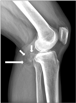
Fig. 1 The plain radiograph of the left knee shows a mass-like lesion containing fat (long arrow) and internal ossifications (short arrows) in the posterior area of the left knee.

Fig. 2 The ultrasound (US) scan of US-guided biopsy reveals a well-defined soft tissue mass and prominent acoustic shadowing caused by internal ossifications.

Fig. 3 Sagittal plane magnetic resonance imaging scans: (A) T1-weighted image, (B) T2-weighted image, (C) fat-suppressed gadolinium-enhanced image, and (D) axial T1-weighted image. The contents of the mass are presumed to be fat (arrows), enhancing fibrous tissue (arrow heads), and ossifications. The tibial nerve (curved arrow) is shown separately.

Fig. 4 Photograph of the popliteal mass. The mass is well-circumscribed and the cut surface is yellow and fatty with traversing whitish fibro-chondroid bands and nodules.
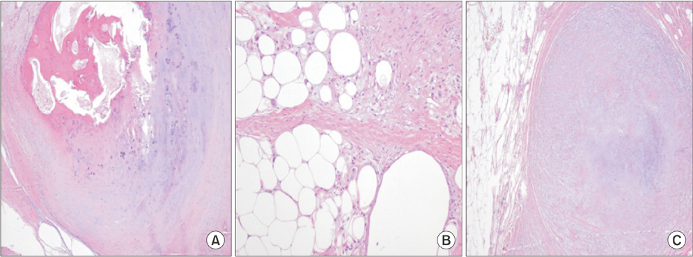
Fig. 5 Histological sections of the tumor specimen show lipoma with cartilaginous and osseous differentiation. (A) Ossification in the chondroid nodule. Lamellar bone with osteoblasts and osteoclasts is being formed in the chondroid nodule (H&E, ×40). (B) Fibroblast proliferation and fibrous band is intimately apposed to the fat necrosis area of lipoma (H&E, ×100). (C) A chondroid nodule in the lipoma. Chondroid matrix and chondrocytes are arising in the fibromyxoid band (H&E, ×40).
Reference
-
1. Baker WM. On the formation of synovial cysts in the leg in connection with disease of the knee-joint. 1877. Clin Orthop Relat Res. 1994; (299):2–10.2. Fritschy D, Fasel J, Imbert JC, Bianchi S, Verdonk R, Wirth CJ. The popliteal cyst. Knee Surg Sports Traumatol Arthrosc. 2006; 14(7):623–628.3. Fiori R, Chiappa R, Gaspari E, Simonetti G. A rare case of popliteal venous aneurysm. Case Rep Med. 2010; 2010:579256.4. Miller TT, Staron RB, Koenigsberg T, Levin TL, Feldman F. MR imaging of Baker cysts: association with internal derangement, effusion, and degenerative arthropathy. Radiology. 1996; 201(1):247–250.5. Sansone V, De Ponti A. Arthroscopic treatment of popliteal cyst and associated intra-articular knee disorders in adults. Arthroscopy. 1999; 15(4):368–372.6. Tseng KF, Hsu HC, Wang FC, Fong YC. Nerve sheath ganglion of the tibial nerve presenting as a Baker's cyst: a case report. Knee Surg Sports Traumatol Arthrosc. 2006; 14(9):880–884.7. Tatari H, Baran O, Lebe B, Kilic S, Manisali M, Havitcioglu H. Pigmented villonodular synovitis of the knee presenting as a popliteal cyst. Arthroscopy. 2000; 16(6):13.8. Shin DS, Kwack BH, Ahn JC. Treatment of synovial sarcoma in popliteal fossa adjacent to tibia. J Korean Bone Joint Tumor Soc. 2007; 13(2):201–206.9. Gru AA, Santa Cruz DJ. Osteochondrolipoma: a subcutaneous lipoma with chondroid and bone differentiation of the chest wall. J Cutan Pathol. 2012; 39(4):461–463.10. Rau T, Soeder S, Olk A, Aigner T. Parosteal lipoma of the thigh with cartilaginous and osseous differentiation: an osteochondrolipoma. Ann Diagn Pathol. 2006; 10(5):279–282.
- Full Text Links
-
- Actions
-
Cited
- CITED
-
- Close
- Share
-
- Similar articles
-
- Tibial Schwannoma Mimicking a Popliteal Cyst
- A Popliteal Cyst Causing Tibial Nerve Entrapment Neuropathy: A Case Report
- Relationship between Popliteal Cyst and the Intra-articular Knee Disorders
- Ruptured Popliteal Cyst Diagnosed by Ultrasound Before Evaluation for Deep Vein Thrombosis
- A Case of Ruptured Popliteal Cyst in Gouty Arthritis
