Tarantula cubensis extract alters the degree of apoptosis and mitosis in canine mammary adenocarcinomas
- Affiliations
-
- 1Department of Obstetrics and Gynecology, Faculty of Veterinary Medicine, University of Ondokuz Mayis, Samsun 55200, Turkey. nilgung@omu.edu.tr
- 2Department of Pathology, Faculty of Veterinary Medicine, University of Ondokuz Mayis, Samsun 55200, Turkey.
- 3Department of Obstetrics and Gynecology, Faculty of Veterinary Medicine, University of Kafkas, Kars 36000, Turkey.
- 4Department of Obstetrics and Gynecology, Faculty of Veterinary Medicine, University of Mehmet Akif Ersoy, Burdur 15030, Turkey.
- 5Department of Obstetrics and Gynecology, Faculty of Veterinary Medicine, University of Dicle, Diyarbakir 21830, Turkey.
- 6Department of Obstetrics and Gynecology, Faculty of Veterinary Medicine, University of Harran, Urfa 63300, Turkey.
- 7Centre for Artificial Insemination and Embryo Transfer, University of Veterinary Medicine Vienna, Vienna A-1210, Austria.
- 8Department of Obstetrics and Gynecology, Faculty of Veterinary Medicine, University of Ankara, Ankara 06100, Turkey.
- KMID: 2164531
- DOI: http://doi.org/10.4142/jvs.2015.16.2.213
Abstract
- In the present study, 13 clinical cases of canine mammary adenocarcinoma were evaluated in order to understand the effect of Tarantula cubensis extract (TCE) on tumor tissue. Punch biopsies were taken from the tumors before treatment with TCE. Subcutaneous injections of TCE were administered three times at weekly intervals (3 mL per dog). Between days 7 and 10 after the third injection, the tumor masses were extirpated by complete unilateral mastectomy. Pre- and post-treatment tumor tissues were immunohistochemically assessed. The expression of B-cell lymphoma 2 (Bcl-2) was found to be higher in pre-treatment compared to post-treatment tissues (p < 0.01) whereas Ki-67 expression was lower in post-treatment tissues (p < 0.01). No significant differences in fibroblast growth factor or vascular endothelial growth factor expression were observed between pre- and post-treatment tissues (p > 0.05). The apoptotic index was determined to be low before treatment and increased during treatment. These results suggest that TCE may be effective for controlling the local growth of canine mammary adenocarcinoma by regulating apoptosis.
Keyword
MeSH Terms
Figure
-
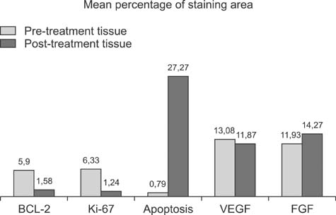
Fig. 1 Mean percentage of areas positive for Bcl-2 (p < 0.01), Ki-67 (p < 0.01), apoptosis (p < 0.01), vascular endothelial growth factor (VEGF; p > 0.05), and fibroblast growth factor (FGF; p > 0.05).
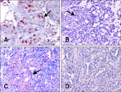
Fig. 2 Immunohistochemical staining for Ki-67 and B-cell lymphoma 2 (Bcl-2). (A) Pre-treatment Ki-67-positive cells (arrow). (B) Post-treatment Ki-67-positive cells (arrow). (C) Pre-treatment Bcl-2-positive cells (arrow). (D) Post-treatment Bcl-2-positive cells. Immunoperoxidase technique with Mayer's haematoxylin counterstaining. Magnification: 240× (A), 180× (B and C), 140× (D).
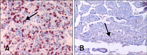
Fig. 3 Pre- and post-treatment terminal deoxynucleotidyl transferase deoxyuridine triphosphate nick end labeling (TUNEL) results. (A) Post-treatment TUNEL-positive apoptotic cells (arrow). (B) Pre-treatment TUNEL-positive apoptotic cells (arrow). TUNEL assay with Mayer's haematoxylin counterstaining, Magnification: 240× (A), 140× (B).
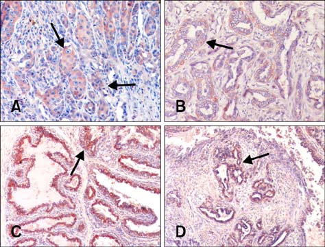
Fig. 4 Immunohistochemical staining for VEGF and FGF. (A) Pre-treatment VEGF-positive cells (arrow). (B) Post-treatment VEGF-positive cells (arrow) (C) Pre-treatment FGF-positive cells (arrow). (D) Post-treatment FGF-positive cells (arrow). Immunoperoxidase technique with Mayer's haematoxylin counterstaining, Magnification: 180× (A and B), 140× (C and D).
Reference
-
1. Abbas AK, Lichtman AH. Cellular and Molecular Immunology. 5th ed. Philadelphia: Saunders;2003. p. 216–240.2. Al-Dissi AN, Haines DM, Singh B, Kidney BA. Immunohistochemical expression of vascular endothelial growth factor and vascular endothelial growth factor receptor-2 in canine simple mammary gland adenocarcinomas. Can Vet J. 2010; 51:1109–1114.3. Alsabeh R, Wilson CS, Ahn CW, Vasef MA, Battifora H. Expression of bcl-2 by breast cancer: a possible diagnostic application. Mod Pathol. 1996; 9:439–444.4. Bamberger ES, Perrett CW. Angiogenesis in epithelian ovarian cancer. Mol Pathol. 2002; 55:348–359.
Article5. Castagnaro M, De Maria R, Bozzetta E, Ru G, Casalone C, Biolatti B, Caramelli M. Ki-67 index as indicator of the post-surgical prognosis in feline mammary carcinomas. Res Vet Sci. 1998; 65:223–226.
Article6. Chen X, Pei L, Zhong Z, Guo J, Zhang Q, Wang Y. Anti-tumor potential of ethanol extract of Curcuma phaeocaulis Valeton against breast cancer cells. Phytomedicine. 2011; 18:1238–1243.
Article7. Daidone MG, Luisi A, Veneroni S, Benini E, Silvestrini R. Clinical studies of Bcl-2 and treatment benefit in breast cancer patients. Endocr Relat Cancer. 1999; 6:61–68.
Article8. Dobson JM, Gorman NT. Cancer Chemotherapy in Small Animal Practice. Oxford: Blackwell Scientific;1993.9. Dole M, Nuñez G, Merchant AK, Maybaum J, Rode CK, Bloch CA, Castle VP. Bcl-2 inhibits chemotherapy-induced apoptosis in neuroblastoma. Cancer Res. 1994; 54:3253–3259.10. Gee JM, Robertson JF, Ellis IO, Willsher P, McClelland RA, Hoyle HB, Kyme SR, Finlay P, Blamey RW, Nicholson RI. Immunocytochemical localization of bcl-2 protein in human breast cancers and its relationship to a series of prognostic markers and response to endocrine therapy. Int J Cancer. 1994; 59:619–628.
Article11. Geraldes M, Gärtner F, Schmitt F. Immunohistochemical study of hormonal receptors and cell proliferation in normal canine mammary glands and spontaneous mammary tumours. Vet Rec. 2000; 146:403–406.
Article12. Gerdes J, Li L, Schlueter C, Duchrow M, Wohlenberg C, Gerlach C, Stahmer I, Kloth S, Brandt E, Flad HD. Immunobiochemical and molecular biologic characterization of the cell proliferation-associated nuclear antigen that is defined by monoclonal antibody Ki-67. Am J Pathol. 1991; 138:867–873.13. Gültiken N, Vural MR. The effect of Tarantula cubensis extract applied in pre and postoperative period of canine mammary tumours. J Istanb Vet Sci. 2007; 2:13–23.14. Halper J. Growth factors as active participants in carcinogenesis: a perspective. Vet Pathol. 2010; 47:77–97.
Article15. Hamilton D. Homeopathic Care for Cats and Dogs: Small Doses for Small Animals. Berkley: North Atlantic Books;1999. p. 237–272.16. Hockenbery D, Nuñez G, Milliman C, Schreiber RD, Korsmeyer SJ. Bcl-2 is an inner mitochondrial membrane protein that blocks programmed cell death. Nature. 1990; 348:334–336.
Article17. Johnston SD, Kustritz MVR, Olson PNS. Canine and Feline Theriogenology. 1st ed. Philadelphia: Saunders;2001. p. 243–256.18. Keen JC, Dixon JM, Miller EP, Cameron DA, Chetty U, Hanby A, Bellamy C, Miller WR. The expression of Ki-S1 and BCL-2 and response to primary tamoxifen therapy in elderly patients with breast cancer. Breast Cancer Res Treat. 1997; 44:123–133.
Article19. Koch H, Stein M. Conservative treatment of neoplasms of the mammary gland in bitches, with the use of Theranekron. Prakt Tierarzt. 1980; 61:429–430.20. Kumar R, Vadlamudi RK, Adam L. Apoptosis in mammary gland and cancer. Endocr Relat Cancer. 2000; 7:257–269.
Article21. Kumaraguruparan R, Prathiba D, Nagini S. Of humans and canines: immunohystochemical analysis of PCNA, Bcl-2, p53, cytokeratin and ER in mammary tumours. Res Vet Sci. 2006; 81:218–224.
Article22. Löhr CV, Teifke JP, Failing K, Weiss E. Characterization of the proliferation state in canine mammary tumors by the standardized AgNOR method with postfixation and immunohistologic detection of Ki-67 and PCNA. Vet Pathol. 1997; 34:212–221.
Article23. MacEwen EG, Withrow SJ. Tumors of the mammary gland. In : Withrow SJ, MacEwen EG, editors. Clinical Veterinary Oncology. Philadelphia: Lippincott;1989. p. 292–304.24. Misdorp W. Tumors of the mammary gland. In : Meuten DJ, editor. Tumors in Domestic Animals. 4th ed. Ames: Iowa State Press;2002. p. 575–606.25. Misdorp W, Else RW, Hellmén E, Lipscomb TP. Histological Classification of Mammary Tumors of the Dog and Cat. 2nd ed. Washigton D.C.: Armed Forces Institute of Pathology;1999. p. 1–59.26. Osborne CA. Generating the diagnosis and prognosis of cancer in geriatric pets. In : Villalobos A, Kaplan L, editors. Canine and Feline Geriatric Oncology: Honoring the Human-Animal Bond. 1st ed. Ames: Blackwell;2007. p. 103–134.27. Owen LN. TNM Classification of Tumours in Domestic Animals. 1st ed. Geneva: World Health Organization;1980.28. Ozmen O, Haligur M, Kocamuftuoglu M. Clinocopathologic and immunohistochemical findings of multiple genital leiomyomas and mammary adenocarcinomas in a bitch. Reprod Domest Anim. 2008; 43:377–381.
Article29. Qiu C, Lin DD, Wang HH, Qiao CH, Wang J, Zhang T. Quantification of VEGF-C expression in canine mammary tumours. Aust Vet J. 2008; 86:279–282.
Article30. Qiu CW, Lin DG, Wang JQ, Li CY, Deng GZ. Expression and significance of PTEN and VEGF in canine mammary gland tumours. Vet Res Commun. 2008; 32:463–472.
Article31. Restucci B, Papparella S, Maiolino P, De Vico G. Expression of vascular endothelial growth factor in canine mammary tumours. Vet Pathol. 2002; 39:488–493.
Article32. Santos AA, Oliveira JT, Lopes CCC, Amorim IF, Vicente CMFB, Gärtner FRM, Matos AJF. Immunohistochemical expression of vascular endothelial growth factor in canine mammary tumours. J Comp Pathol. 2010; 143:268–275.
Article33. Sarli G, Preziosi R, Benazzi C, Castellani G, Marcato PS. Prognostic value of histologic stage and proliferative activity in canine malignant mammary tumors. J Vet Diagn Invest. 2002; 14:25–34.
Article34. Tewari M, Pradhan S, Singh U, Singh TB, Shukla HS. Assesment of predictive markers of response to neoadjuvant chemotherapy in breast cancer. Asian J Surg. 2010; 33:157–167.
Article35. Thuróczy J, Reisvaag GJ, Perge E, Tibold A, Szilágyi J, Balogh L. Immunohistochemical detection of progesteron and cellular proliferation in canine mammary tumours. J Comp Pathol. 2007; 137:122–129.
Article36. Weidner N. Tumoural vascularity as a prognostic factor in cancer patients: the evidence continues to grow. J Pathol. 1998; 184:119–122.
Article37. Yang WY, Liu CH, Chang CJ, Lee CC, Chang KJ, Lin CT. Proliferative activity, apoptosis and expression of oestrogen receptor and Bcl-2 oncoprotein in canine mammary gland tumours. J Comp Pathol. 2006; 134:70–79.
Article38. Zuccari DA, Santana AE, Cury PM, Cordeiro JA. Immunocytochemical study of Ki-67 as a prognostic marker in canine mammary neoplasia. Vet Clin Pathol. 2004; 33:23–28.
Article
- Full Text Links
-
- Actions
-
Cited
- CITED
-
- Close
- Share
-
- Similar articles
-
- Mutations of p53 Tumor Suppressor Gene in Spontaneous Canine Mammary Tumors
- Alpha basic crystallin expression in canine mammary tumors
- Co-Occurrence of Two Phylogenetic Clades of Pseudoperonospora cubensis, the Causal Agent of Downy Mildew Disease, on Oriental Pickling Melon
- In vitro evaluation of the antitumor activity of axitinib in canine mammary gland tumor cell lines
- The Effect of Paclitaxel(Taxol) on the Radiation in the Rat Liver
