Transvaginal/transrectal ultrasound for assessing myometrial invasion in endometrial cancer: a comparison of six different approaches
- Affiliations
-
- 1Department of Obstetrics and Gynecology, Clinica Universidad de Navarra, University of Navarra School of Medicine, Pamplona, Spain. jlalcazar@unav.es
- 2Department of Obstetrics and Gynecology, Hospital de Cruces, Bilbao, Spain.
- 3Department of Obstetrics and Gynecology, Hospital de la Arrixaca, Murcia, Spain.
- KMID: 2160806
- DOI: http://doi.org/10.3802/jgo.2015.26.3.201
Abstract
OBJECTIVE
To compare the diagnostic performance of six different approaches for assessing myometrial infiltration using ultrasound in women with carcinoma of the corpus uteri.
METHODS
Myometrial infiltration was assessed by two-dimensional (2D) transvaginal or transrectal ultrasound in 169 consecutive women with well (G1) or moderately (G2) differentiated endometrioid type endometrial carcinoma. In 74 of these women three-dimensional (3D) ultrasound was also performed. Six different techniques for myometrial infiltration assessment were evaluated. The impression of examiner and Karlsson's criteria were assessed prospectively. Endometrial thickness, tumor/uterine 3D volume ratio, tumor distance to myometrial serosa (TDS), and van Holsbeke's subjective model were assessed retrospectively. All subjects underwent surgical staging within 1 week after ultrasound evaluation. Definitive histopathological data regarding myometrial infiltration was used as gold standard. Sensitivity and specificity for all approaches were calculated and compared using McNemar test.
RESULTS
The impression of examiner and subjective model performed similarly (sensitivity 79.5% and 80.5%, respectively; specificity 89.6% and 90.3%, respectively). Both methods had significantly better sensitivity than Karlsson's criteria (sensitivity 31.8%, p<0.05) and endometrial thickness (sensitivity 47.7%, p<0.05), and better specificity than tumor/uterine volume ratio (specificity 28.3%, p<0.05) and TDS (specificity 41.5%, p<0.05).
CONCLUSION
Subjective impression seems to be the best approach for assessing myometrial infiltration in G1 or G2 endometrioid type endometrial cancer by transvaginal or transrectal ultrasound. The use of mathematical models and other objective 2D and 3D measurement techniques do not improve diagnostic performance.
MeSH Terms
-
Adult
Aged
Aged, 80 and over
Carcinoma, Endometrioid/pathology/*ultrasonography
Endometrial Neoplasms/pathology/*ultrastructure
Female
Humans
Imaging, Three-Dimensional
Middle Aged
Models, Theoretical
Myometrium/pathology/ultrasonography
Neoplasm Invasiveness/pathology/*ultrasonography
Prospective Studies
Retrospective Studies
Tumor Burden
Figure
-
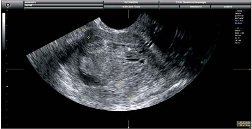
Fig. 1 Transvaginal ultrasound showing measurement of tumor/anteroposterior uterine diameter ratio as proposed by Karlsson. In this case the ratio is ≥50% indicating myometrial infiltration of ≥50%.
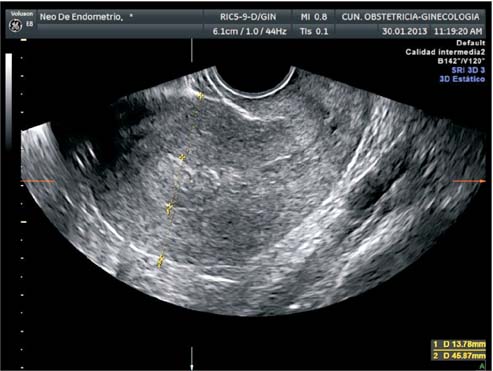
Fig. 2 Transvaginal ultrasound showing measurement of tumor/anteroposterior uterine diameter ratio as proposed by Karlsson. In this case the ratio is <50% indicating myometrial infiltration of <50%.
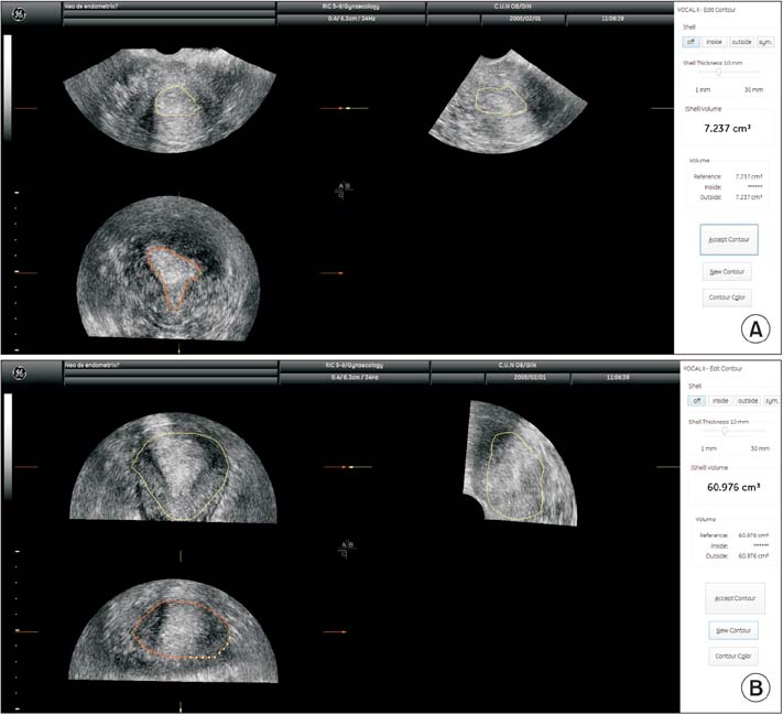
Fig. 3 Three-dimensional ultrasound estimation of tumor (A) and uterine volumes (B). In this case the ratio is 0.118, indicating myometrial infiltration of <50%.
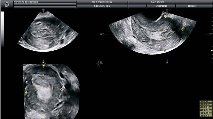
Fig. 4 Three-dimensional ultrasound estimation of shortest tumor distance to serosa (TDS). In this case TDS is 10.5 mm, indicating myometrial infiltration of <50%.
Cited by 1 articles
-
Falsely Elevated Postvoid Residual Urine Volume in Uterine Myoma
Tae Hee Kim, Hyo Sang Kim, Jung Wook Park, Oh Kyung Lim, Ki Deok Park, Ju Kang Lee
Ann Rehabil Med. 2017;41(2):332-336. doi: 10.5535/arm.2017.41.2.332.
Reference
-
1. Creasman WT, Morrow CP, Bundy BN, Homesley HD, Graham JE, Heller PB. A Gynecologic Oncology Group Study. Surgical pathologic spread patterns of endometrial cancer. Cancer. 1987; 60:8 Suppl. 2035–2041.2. Morrow CP, Bundy BN, Kurman RJ, Creasman WT, Heller P, Homesley HD, et al. Relationship between surgical-pathological risk factors and outcome in clinical stage I and II carcinoma of the endometrium: a Gynecologic Oncology Group study. Gynecol Oncol. 1991; 40:55–65.3. Bogani G, Dowdy SC, Cliby WA, Ghezzi F, Rossetti D, Mariani A. Role of pelvic and para-aortic lymphadenectomy in endometrial cancer: current evidence. J Obstet Gynaecol Res. 2014; 40:301–311.4. SGO Clinical Practice Endometrial Cancer Working Group, Burke WM, Orr J, Leitao M, Salom E, Gehrig P, et al. Endometrial cancer: a review and current management strategies: part I. Gynecol Oncol. 2014; 134:385–392.5. Kumar S, Medeiros F, Dowdy SC, Keeney GL, Bakkum-Gamez JN, Podratz KC, et al. A prospective assessment of the reliability of frozen section to direct intraoperative decision making in endometrial cancer. Gynecol Oncol. 2012; 127:525–531.6. Cacciatore B, Lehtovirta P, Wahlstrom T, Ylanen K, Ylostalo P. Contribution of vaginal scanning to sonographic evaluation of endometrial cancer invasion. Acta Oncol. 1989; 28:585–588.7. Cagnazzo G, D'Addario V, Martinelli G, Lastilla G. Depth of myometrial invasion in endometrial cancer: preoperative assessment by transvaginal ultrasonography and magnetic resonance imaging. Ultrasound Obstet Gynecol. 1992; 2:40–43.8. Osmers RG, Osmers M, Kuhn W. Prognostic value of transvaginal sonography in asymptomatic endometrial cancers. Ultrasound Obstet Gynecol. 1995; 6:103–107.9. van Doorn HC, van der Zee AG, Peeters PH, Kroeks MV, van Eijkeren MA. Preoperative selection of patients with low-stage endometrial cancer at high risk of pelvic lymph node metastases. Int J Gynecol Cancer. 2002; 12:144–148.10. Savelli L, Ceccarini M, Ludovisi M, Fruscella E, De Iaco PA, Salizzoni E, et al. Preoperative local staging of endometrial cancer: transvaginal sonography vs. magnetic resonance imaging. Ultrasound Obstet Gynecol. 2008; 31:560–566.11. Antonsen SL, Jensen LN, Loft A, Berthelsen AK, Costa J, Tabor A, et al. MRI, PET/CT and ultrasound in the preoperative staging of endometrial cancer: a multicenter prospective comparative study. Gynecol Oncol. 2013; 128:300–308.12. Gordon AN, Fleischer AC, Reed GW. Depth of myometrial invasion in endometrial cancer: preoperative assessment by transvaginal ultrasonography. Gynecol Oncol. 1990; 39:321–327.13. Karlsson B, Norstrom A, Granberg S, Wikland M. The use of endovaginal ultrasound to diagnose invasion of endometrial carcinoma. Ultrasound Obstet Gynecol. 1992; 2:35–39.14. Fotopoulou C, Sehouli J, Schefold JC, Wolf C, Denkert C, Lichtenegger W, et al. Preoperative transvaginal ultrasound (TVS) in the description of pelvic tumor spread in endometrial cancer: results of a prospective study. Anticancer Res. 2008; 28(4C):2453–2458.15. De Smet F, De Brabanter J, Van den Bosch T, Pochet N, Amant F, Van Holsbeke C, et al. New models to predict depth of infiltration in endometrial carcinoma based on transvaginal sonography. Ultrasound Obstet Gynecol. 2006; 27:664–671.16. Van Holsbeke C, Ameye L, Testa AC, Mascilini F, Lindqvist P, Fischerova D, et al. Development and external validation of new ultrasound-based mathematical models for preoperative prediction of high-risk endometrial cancer. Ultrasound Obstet Gynecol. 2014; 43:586–595.17. Alcazar JL, Galvan R, Albela S, Martinez S, Pahisa J, Jurado M, et al. Assessing myometrial infiltration by endometrial cancer: uterine virtual navigation with three-dimensional US. Radiology. 2009; 250:776–783.18. Mascilini F, Testa AC, Van Holsbeke C, Ameye L, Timmerman D, Epstein E. Evaluating myometrial and cervical invasion in women with endometrial cancer: comparing subjective assessment with objective measurement techniques. Ultrasound Obstet Gynecol. 2013; 42:353–358.19. Alcazar JL, Pineda L, Caparros M, Utrilla-Layna J, Juez L, Minguez JA, et al. Transvaginal/transrectal ultrasound for preoperative identification of high-risk cases in well or moderately differentiated endometrioid carcinoma of the endometrium. Ultrasound Obstet Gynecol. 2015; 05. 29. [Epub]. DOI: 10.1002/uog.14912.20. Lewin SN. Revised FIGO staging system for endometrial cancer. Clin Obstet Gynecol. 2011; 54:215–218.21. Eriksson LS, Lindqvist PG, Floter Radestad A, Dueholm M, Fischerova D, Franchi D, et al. Transvaginal ultrasound assessment of myometrial and cervical stromal invasion in women with endometrial cancer: interobserver reproducibility among ultrasound experts and gynecologists. Ultrasound Obstet Gynecol. 2015; 45:476–482.22. Prompeler HJ, Madjar H, du Bois A, Lattermann U, Wilhelm C, Kommoss F, et al. Transvaginal sonography of myometrial invasion depth in endometrial cancer. Acta Obstet Gynecol Scand. 1994; 73:343–346.23. Weber G, Merz E, Bahlmann F, Mitze M, Weikel W, Knapstein PG. Assessment of myometrial infiltration and preoperative staging by transvaginal ultrasound in patients with endometrial carcinoma. Ultrasound Obstet Gynecol. 1995; 6:362–367.24. Gabrielli S, Marabini A, Bevini M, Linsalata I, Falco P, Milano V, et al. Transvaginal sonography vs. hysteroscopy in the preoperative staging of endometrial carcinoma. Ultrasound Obstet Gynecol. 1996; 7:443–446.25. Sawicki W, Spiewankiewicz B, Stelmachow J, Cendrowski K. The value of ultrasonography in preoperative assessment of selected prognostic factors in endometrial cancer. Eur J Gynaecol Oncol. 2003; 24:293–298.26. Akbayir O, Corbacioglu A, Numanoglu C, Guleroglu FY, Ulker V, Akyol A, et al. Preoperative assessment of myometrial and cervical invasion in endometrial carcinoma by transvaginal ultrasound. Gynecol Oncol. 2011; 122:600–603.
- Full Text Links
-
- Actions
-
Cited
- CITED
-
- Close
- Share
-
- Similar articles
-
- Diagnostic value of transvaginal contrast-enhanced ultrasound in identifying benign and malignant endometrial lesions and assessing myometrial invasion
- Usefulness of Sonohysterography in the Assessment of Myometrial Invasion in Endometrial Carcinoma
- Comparison of MRI's Prediction of Myometrial, Cervical Invasion and Surgical Staging in Endometrial Carcinoma
- Transrectal ultrasonography: evaluation of bladder wall inversion in cervical cancer
- Usefulness of Sonohy sterography in Differentiating Endometrial Cancer from Endometrial Hyperp lasia
