A Pleural Loose Body Mimicking a Pleural Tumor: A Case Report
- Affiliations
-
- 1Department of Radiology, School of Medicine, Ewha Womans University, Seoul 07985, Korea. yookkim@ewha.ac.kr
- 2Division of Pulmonology and Critical Care Medicine in Department of Internal Medicine, School of Medicine, Ewha Womans University, Seoul 07985, Korea.
- 3Department of Thoracic and Cardiovascular Surgery, School of Medicine, Ewha Womans University, Seoul 07985, Korea.
- 4Department of Pathology, School of Medicine, Ewha Womans University, Seoul 07985, Korea.
- KMID: 2160784
- DOI: http://doi.org/10.3348/kjr.2015.16.5.1163
Abstract
- We present a rare case of a pleural loose body, thought to be a pedunculated pleural tumor, found incidentally in a 58-year-old female. Computed tomography showed a non-enhancing mass, which migrated along the mediastinum and paravertebral area. Thoracoscopic surgery revealed a 4 cm, soap-like mass that was found to be a fibrin body consisting of hyalinized collagen histopathologically. Mobility and the lack of contrast enhancement of a pleural mass are important clues to diagnosing this benign condition.
MeSH Terms
Figure
-
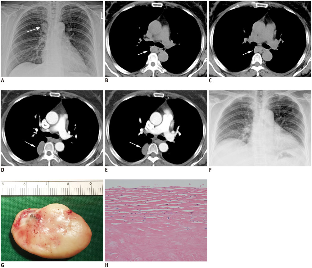
Fig. 1 58-year-old woman with pleural fibrin body. A. Posteroanterior chest radiograph shows right paratracheal mass (arrow). B, C. Pre-contrast CT demonstrates soft-tissue density mass in subcarinal area (arrows). Attenuation of mass was 42-44 Hounsfield units (HU). D, E. On postcontrast CT, mass is observed in right paravertebral area (arrows). Mass did not show contrast enhancement and had attenuation value around 44 HU. F. On preoperative chest radiograph obtained 1 month after initial radiograph, no mass is observed in right paratracheal area. Increased opacity in subcarinal area suggests migration of previous right paratracheal mass to subcarinal area. G. Gross tumor specimen is whitish "soap-like" mass with smooth surface. H. In high-power view, surface of tumor shows scattered chronic inflammatory cells and characteristic "basket-weave" configuration of laminated, hypocellular mature collagen (hematoxylin-eosin stain, × 200 magnification).
Reference
-
1. Bolca C, Trahan S, Frechette E. Intrapleural thoracolithiasis: a rare intrathoracic pearl-like lesion. Thorac Cardiovasc Surg. 2011; 59:445–446.2. Peungjesada S, Gupta P, Mottershaw AM. Thoracolithiasis: a case report. Clin Imaging. 2012; 36:228–230.3. Kinoshita F, Saida Y, Okajima Y, Honda S, Sato T, Hayashibe A, et al. Thoracolithiasis: 11 cases with a calcified intrapleural loose body. J Thorac Imaging. 2010; 25:64–67.4. Spitz WU, Taff ML. Intrapleural golf ball size loose body. An incidental finding at autopsy. Am J Forensic Med Pathol. 1985; 6:329–331.5. Kawanami T. [Post-thoracotomy "fibrin body": 16 year follow-up of a case (author's transl)]. Rinsho Hoshasen. 1980; 25:863–866.6. Euphrat EJ, Beck E. Fibrin body following traumatic pneumothorax; a problem in differential diagnosis of a nodular pulmonary density. Am J Roentgenol Radium Ther Nucl Med. 1955; 74:86–89.7. Dias AR, Zerbini EJ, Curi N. Pleural stone. A case report. J Thorac Cardiovasc Surg. 1968; 56:120–122.8. Iwasaki T, Nakagawa K, Katsura H, Ohse N, Nagano T, Kawahara K. Surgically removed thoracolithiasis: report of two cases. Ann Thorac Cardiovasc Surg. 2006; 12:279–282.9. Tanaka D, Niwatsukino H, Fujiyoshi F, Nakajo M. Thoracolithiasis--a mobile calcified nodule in the intrathoracic space: radiographic, CT, and MRI findings. Radiat Med. 2002; 20:131–133.10. Kosaka S, Kondo N, Sakaguchi H, Kitano T, Harada T, Nakayama K. Thoracolithiasis. Jpn J Thorac Cardiovasc Surg. 2000; 48:318–321.
- Full Text Links
-
- Actions
-
Cited
- CITED
-
- Close
- Share
-
- Similar articles
-
- Imaging Features of Various Benign and Malignant Tumors and Tumorlike Conditions of the Pleura: A Pictorial Review
- Pleural Calcification as a Manifestation of Paragonimiasis: A Report of Two Cases
- A Case of a Huge Mass Due to Pleural Metastasis and Management of Dyspnea in a Patient with Terminal Breast Cancer
- Diagnostic Tools of Pleural Effusion
- Intrathoracic Desmoid Tumor Mimicking Pleural Mass: A Case Report
