Deltoid Ligament and Tibiofibular Syndesmosis Injury in Chronic Lateral Ankle Instability: Magnetic Resonance Imaging Evaluation at 3T and Comparison with Arthroscopy
- Affiliations
-
- 1Department of Radiology, Eulji Hospital, Eulji University, Seoul 01830, Korea. mdcys0128@hanmail.net
- 2Department of Orthopedic Surgery, Eulji Hospital, Eulji University, Seoul 01830, Korea.
- KMID: 2160777
- DOI: http://doi.org/10.3348/kjr.2015.16.5.1096
Abstract
OBJECTIVE
To evaluate the prevalence of deltoid ligament and distal tibiofibular syndesmosis injury on 3T magnetic resonance imaging (MRI) in patients with chronic lateral ankle instability (CLAI).
MATERIALS AND METHODS
Fifty patients (mean age, 35 years) who had undergone preoperative 3T MRI and surgical treatment for CLAI were enrolled. The prevalence of deltoid ligament and syndesmosis injury were assessed. The complexity of lateral collateral ligament complex (LCLC) injury was correlated with prevalence of deltoid or syndesmosis injuries. The diagnostic accuracy of ankle ligament imaging at 3T MRI was analyzed using arthroscopy as a reference standard.
RESULTS
On MRI, deltoid ligament injury was identified in 18 (36%) patients as follows: superficial ligament alone, 9 (50%); deep ligament alone 2 (11%); and both ligaments 7 (39%). Syndesmosis abnormality was found in 21 (42%) patients as follows: anterior inferior tibiofibular ligament (AITFL) alone, 19 (90%); and AITFL and interosseous ligament, 2 (10%). There was no correlation between LCLC injury complexity and the prevalence of an accompanying deltoid or syndesmosis injury on both MRI and arthroscopic findings. MRI sensitivity and specificity for detection of deltoid ligament injury were 84% and 93.5%, and those for detection of syndesmosis injury were 91% and 100%, respectively.
CONCLUSION
Deltoid ligament or syndesmosis injuries were common in patients undergoing surgery for CLAI, regardless of the LCLC injury complexity. 3T MRI is helpful for the detection of all types of ankle ligament injury. Therefore, careful interpretation of pre-operative MRI is essential.
Keyword
MeSH Terms
Figure
-
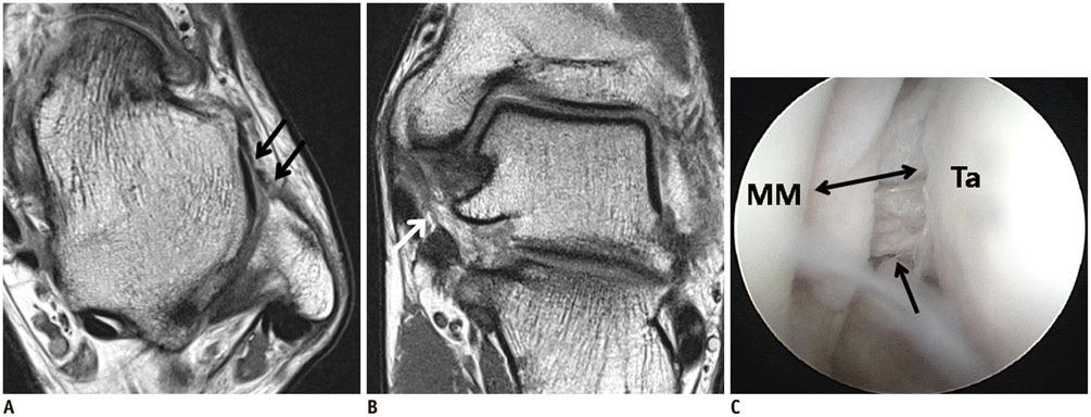
Fig. 1 21-year-old man with chronic tear of anterior talofibular ligament (ATFL) and deltoid ligament. A. Axial proton-weighted turbo spin echo (TSE) magnetic resonance (MR) image showing torn ATFL (arrows). B. Oblique coronal proton-weighted TSE MR image showing loose, thin superficial deltoid ligament (arrow). Deep posterior tibiotalar ligament is thickened with loss of striation. C. On arthroscopy, injury of superficial (not visible on this figure) and deep deltoid ligament (arrow) with widening of medial mortise (double arrow) were seen. Patient underwent modified Broström operation and repair of superficial deltoid ligament. MM = medial malleolus, Ta = medial talus
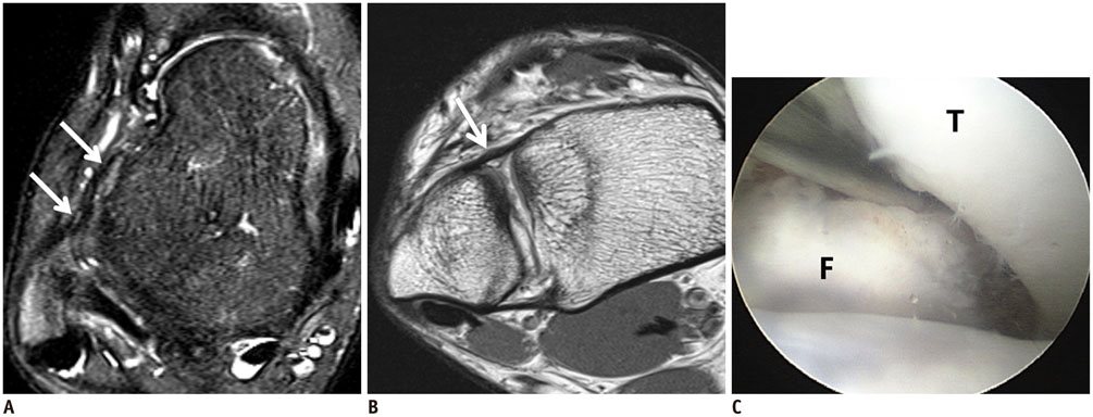
Fig. 2 27-year-old man with chronic injuries of anterior talofibular ligament (ATFL) and anterior inferior tibiofibular ligament (AITFL). A. Axial fat suppression T2-weighted turbo spin echo (TSE) magnetic resonance (MR) image shows very loose and irregular ATFL (arrows). B. Axial oblique proton-weighted TSE MR image shows irregular and loose AITFL (arrow). C. On arthroscopic image, there are obturator insertion and widening of distal tibiofibular joint (> 4 mm). Patient underwent modified Broström operation and reconstruction of AITFL using anchors. F = distal fibula, T = distal tibia
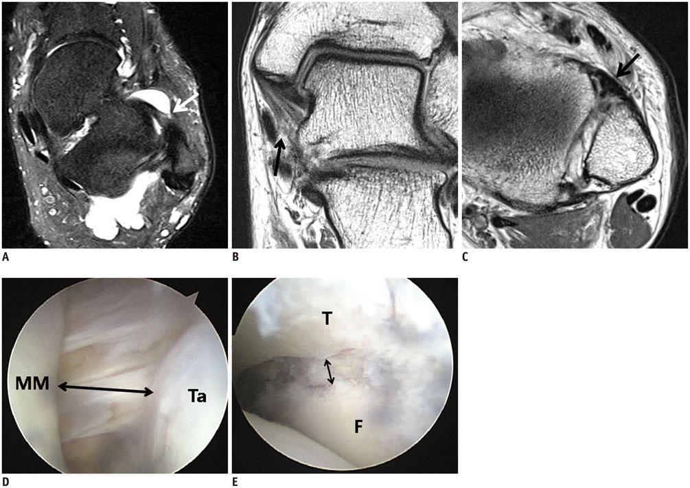
Fig. 3 48-year-old man with chronic injuries to anterior talofibular ligament (ATFL), superficial deltoid ligament, and anterior inferior tibiofibular ligament (AITFL). A. Axial T2-weighted fat-suppression turbo spin echo (TSE) magnetic resonance (MR) image shows hyperintensity of ATFL, with partial discontinuity (arrow). B. Coronal oblique proton-weighted TSE MR image shows hyperintensity of superficial deltoid ligament, with partial discontinuity (arrow). C. Axial proton-weighted TSE MR image shows thickened AITFL (arrow). D, E. Arthroscopic images show loose and partially torn superficial deltoid ligament, with widening of medial mortise (double arrow, D), and widening of distal tibiofibular joint (double arrow, E). Patient underwent modified Broström operation, repair of superficial deltoid ligament, and reconstruction of AITFL using anchors. F = distal fibula, MM = medial malleolus, T = distal tibia, Ta = medial talus
Reference
-
1. Chan KW, Ding BC, Mroczek KJ. Acute and chronic lateral ankle instability in the athlete. Bull NYU Hosp Jt Dis. 2011; 69:17–26.2. Garrick JG. The frequency of injury, mechanism of injury, and epidemiology of ankle sprains. Am J Sports Med. 1977; 5:241–242.3. Cass JR, Morrey BF. Ankle instability: current concepts, diagnosis, and treatment. Mayo Clin Proc. 1984; 59:165–170.4. Alparslan L, Chiodo CP. Lateral ankle instability: MR imaging of associated injuries and surgical treatment procedures. Semin Musculoskelet Radiol. 2008; 12:346–358.5. Ferkel RD, Chams RN. Chronic lateral instability: arthroscopic findings and long-term results. Foot Ankle Int. 2007; 28:24–31.6. Joshy S, Abdulkadir U, Chaganti S, Sullivan B, Hariharan K. Accuracy of MRI scan in the diagnosis of ligamentous and chondral pathology in the ankle. Foot Ankle Surg. 2010; 16:78–80.7. Crim JR, Beals TC, Nickisch F, Schannen A, Saltzman CL. Deltoid ligament abnormalities in chronic lateral ankle instability. Foot Ankle Int. 2011; 32:873–878.8. Milz P, Milz S, Steinborn M, Mittlmeier T, Putz R, Reiser M. Lateral ankle ligaments and tibiofibular syndesmosis. 13-MHz high-frequency sonography and MRI compared in 20 patients. Acta Orthop Scand. 1998; 69:51–55.9. Schneck CD, Mesgarzadeh M, Bonakdarpour A. MR imaging of the most commonly injured ankle ligaments. Part II. Ligament injuries. Radiology. 1992; 184:507–512.10. Kanamoto T, Shiozaki Y, Tanaka Y, Yonetani Y, Horibe S. The use of MRI in pre-operative evaluation of anterior talofibular ligament in chronic ankle instability. Bone Joint Res. 2014; 3:241–245.11. Oae K, Takao M, Uchio Y, Ochi M. Evaluation of anterior talofibular ligament injury with stress radiography, ultrasonography and MR imaging. Skeletal Radiol. 2010; 39:41–47.12. Parisien JS, Shereff MJ. The role of arthroscopy in the diagnosis and treatment of disorders of the ankle. Foot Ankle. 1981; 2:144–149.13. James PT, Peter DL. Recent advances in ankle arthroscopic techniques. In : Parisien JS, editor. Current Techniques in Arthroscopy. 3rd ed. New York: Thieme;1998. p. 182–194.14. Hintermann B, Boss A, Schäfer D. Arthroscopic findings in patients with chronic ankle instability. Am J Sports Med. 2002; 30:402–409.15. Hintermann B, Valderrabano V, Boss A, Trouillier HH, Dick W. Medial ankle instability: an exploratory, prospective study of fifty-two cases. Am J Sports Med. 2004; 32:183–190.16. Han SH, Lee JW, Kim S, Suh JS, Choi YR. Chronic tibiofibular syndesmosis injury: the diagnostic efficiency of magnetic resonance imaging and comparative analysis of operative treatment. Foot Ankle Int. 2007; 28:336–342.17. Takao M, Ochi M, Oae K, Naito K, Uchio Y. Diagnosis of a tear of the tibiofibular syndesmosis. The role of arthroscopy of the ankle. J Bone Joint Surg Br. 2003; 85:324–329.18. Ogilvie-Harris DJ, Reed SC. Disruption of the ankle syndesmosis: diagnosis and treatment by arthroscopic surgery. Arthroscopy. 1994; 10:561–568.19. Ferran NA, Oliva F, Maffulli N. Ankle instability. Sports Med Arthrosc. 2009; 17:139–145.20. Karlsson J, Lansinger O. Lateral instability of the ankle joint. Clin Orthop Relat Res. 1992; 276:253–261.21. van Rijn RM, van Os AG, Bernsen RM, Luijsterburg PA, Koes BW, Bierma-Zeinstra SM. What is the clinical course of acute ankle sprains? A systematic literature review. Am J Med. 2008; 121:324–331.e6.22. Harrington KD. Degenerative arthritis of the ankle secondary to long-standing lateral ligament instability. J Bone Joint Surg Am. 1979; 61:354–361.23. Hertel J. Functional Anatomy, Pathomechanics, and Pathophysiology of Lateral Ankle Instability. J Athl Train. 2002; 37:364–375.24. Attarian DE, McCrackin HJ, DeVito DP, McElhaney JH, Garrett WE Jr. Biomechanical characteristics of human ankle ligaments. Foot Ankle. 1985; 6:54–58.25. Lee KM, Chung CY, Kwon SS, Chung MK, Won SH, Lee SY, et al. Relationship between stress ankle radiographs and injured ligaments on MRI. Skeletal Radiol. 2013; 42:1537–1542.26. Miller CD, Shelton WR, Barrett GR, Savoie FH, Dukes AD. Deltoid and syndesmosis ligament injury of the ankle without fracture. Am J Sports Med. 1995; 23:746–750.27. McCollum GA, van den Bekerom MP, Kerkhoffs GM, Calder JD, van Dijk CN. Syndesmosis and deltoid ligament injuries in the athlete. Knee Surg Sports Traumatol Arthrosc. 2013; 21:1328–1337.28. Hua Y, Chen S, Li Y, Chen J, Li H. Combination of modified Broström procedure with ankle arthroscopy for chronic ankle instability accompanied by intra-articular symptoms. Arthroscopy. 2010; 26:524–528.29. Hopkinson WJ, St Pierre P, Ryan JB, Wheeler JH. Syndesmosis sprains of the ankle. Foot Ankle. 1990; 10:325–330.30. Teramoto A, Kura H, Uchiyama E, Suzuki D, Yamashita T. Three-dimensional analysis of ankle instability after tibiofibular syndesmosis injuries: a biomechanical experimental study. Am J Sports Med. 2008; 36:348–352.31. Chandnani VP, Harper MT, Ficke JR, Gagliardi JA, Rolling L, Christensen KP, et al. Chronic ankle instability: evaluation with MR arthrography, MR imaging, and stress radiography. Radiology. 1994; 192:189–194.32. Cha SD, Kim HS, Chung ST, Yoo JH, Park JH, Kim JH, et al. Intra-articular lesions in chronic lateral ankle instability: comparison of arthroscopy with magnetic resonance imaging findings. Clin Orthop Surg. 2012; 4:293–299.
- Full Text Links
-
- Actions
-
Cited
- CITED
-
- Close
- Share
-
- Similar articles
-
- Comparison of Clinical Results between Deltoid Ligament Augmentation and Syndesmosis Screw Fixation in Bimalleolar Equivalent Fracture
- Diagnosis of Lateral Ankle Ligament Injury in the Evaluation of Chronic Lateral Ankle Instability
- Ankle Syndesmotic Injury
- Treatement of Diastasis of the Distal Tibiofibular Syndesmosis
- Diagnosis and Management of Suspected Deltoid Injury
