Myocardial Contrast Defect Associated with Thrombotic Coronary Occlusion: Pre-Autopsy Diagnosis of a Cardiac Death with Post-Mortem CT Angiography
- Affiliations
-
- 1Department of Radiology, Soonchunhyang University Hospital, Bucheon 14584, Korea. acarad@naver.com
- 2Department of Forensic Medicine, National Forensic Service, Wonju 26460, Korea.
- KMID: 2160769
- DOI: http://doi.org/10.3348/kjr.2015.16.5.1024
Abstract
- We report the case of a female who died of suspected acute myocardial infarction. Post-mortem CT angiography (PMCTA) was performed with intravascular contrast infusion before the standard autopsy, and it successfully demonstrated the complete thrombotic occlusion of a coronary artery and also a corresponding perfusion defect on myocardium. We herein describe the PMCTA findings of a cardiac death with special emphasis on the potential benefits of this novel CT technique in forensic practice.
MeSH Terms
Figure
-
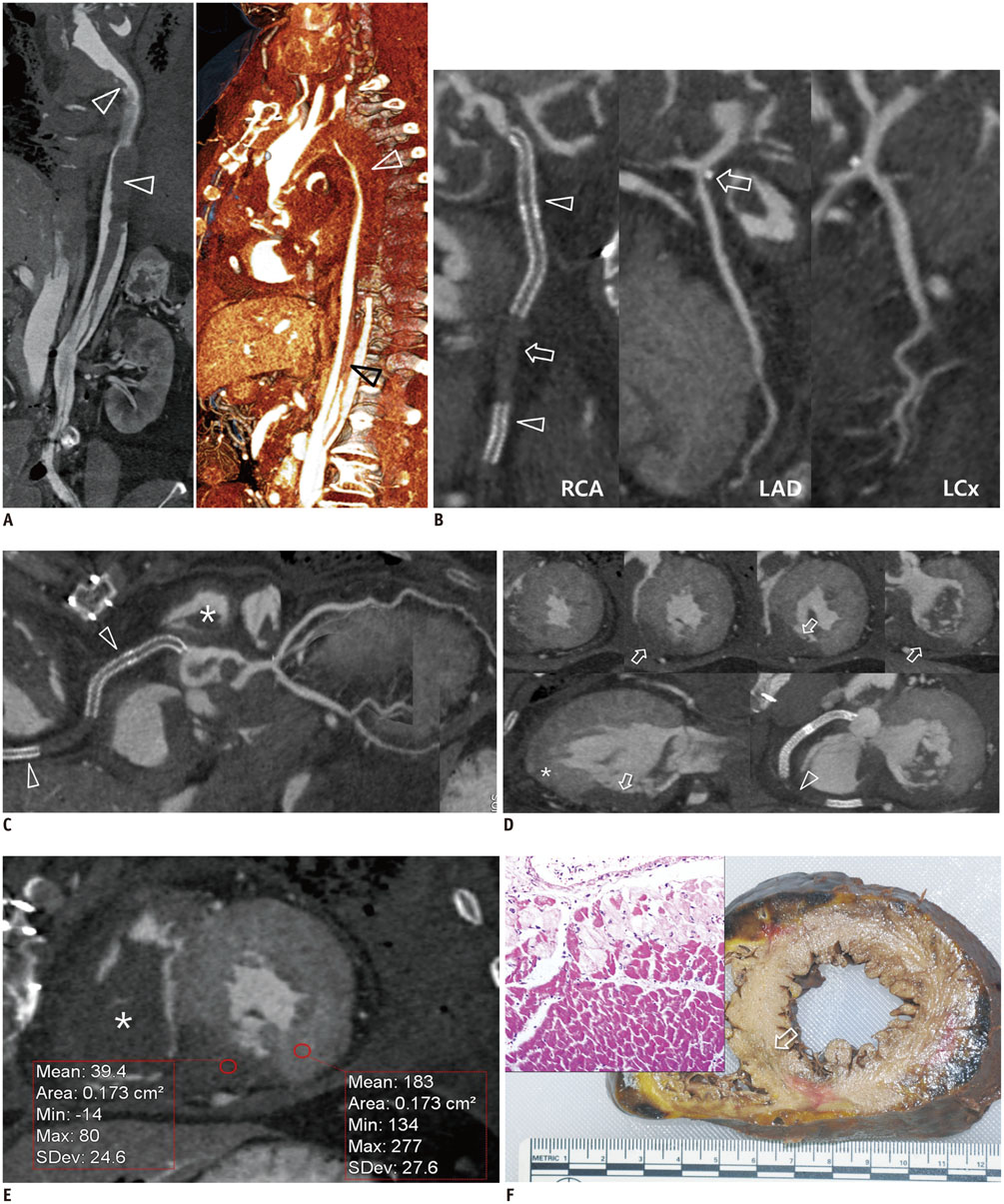
Fig. 1 Post-mortem CT angiography and pathologic specimen of 59-year-old female who died of suspected myocardial infarction. A. Curved multiplanar reformation (MPR, left) and three-dimensional volume rendering image (right) demonstrate aortic dissection with intimo-medial flap and thrombosed false lumen (arrowheads) from aortic arch extending to left common iliac artery. Autopsy confirmed ascending aortic replacement due to previous type A aortic dissection. B, C. Curved MPR (B) and medial axial reformat (C) images of coronary tree show total thrombotic occlusion of stents (arrowheads) in right coronary artery (RCA) and RCA itself (arrow), and also depict focal mixed plaque (arrow) in proximal left anterior descending artery (LAD) and normal left circumflex artery (LCx). Note postmortem clots (*) in pulmonary artery. D. Multiple short-axis views of left ventricle (LV) show localized transmural perfusion defects (arrows) in inferior and inferoseptal walls of mid and basal LV, sharply demarcated from normal myocardium showing diffuse contrast enhancement (upper). Perfusion defects correspond to thrombosed RCA (arrowhead) territory with apical sparing (*) clearly depicted on two-chamber view (lower column). E. Density measurements in short axis image demonstrate considerable contrast enhancement in normal myocardium, that can be attributed to compact filling of oily contrast agent in micro-capillary system through patent LAD and LCx coronary arteries. Note huge postmortem clot (*) in right ventricle. F. Gross and microscopic specimens of heart. Cut surface shows subtle mottling with yellow-tan softening in damaged inferoseptal wall (arrow), which has typical microscopic changes of coagulation necrosis with focal interstitial infiltrate of neutrophils, indicative of acute myocardial infarction (left upper). LAD = left anterior descending artery, LCx = left circumflex artery
Reference
-
1. Bastarrika G, Lee YS, Huda W, Ruzsics B, Costello P, Schoepf UJ. CT of coronary artery disease. Radiology. 2009; 253:317–338.2. Leth PM. Computerized tomography used as a routine procedure at postmortem investigations. Am J Forensic Med Pathol. 2009; 30:219–222.3. Dirnhofer R, Jackowski C, Vock P, Potter K, Thali MJ. VIRTOPSY: minimally invasive, imaging-guided virtual autopsy. Radiographics. 2006; 26:1305–1333.4. Grabherr S, Djonov V, Yen K, Thali MJ, Dirnhofer R. Postmortem angiography: review of former and current methods. AJR Am J Roentgenol. 2007; 188:832–838.5. Grabherr S, Doenz F, Steger B, Dirnhofer R, Dominguez A, Sollberger B, et al. Multi-phase post-mortem CT angiography: development of a standardized protocol. Int J Legal Med. 2011; 125:791–802.6. Michaud K, Grabherr S, Doenz F, Mangin P. Evaluation of postmortem MDCT and MDCT-angiography for the investigation of sudden cardiac death related to atherosclerotic coronary artery disease. Int J Cardiovasc Imaging. 2012; 28:1807–1822.7. Roberts IS, Benamore RE, Peebles C, Roobottom C, Traill ZC. Technical report: diagnosis of coronary artery disease using minimally invasive autopsy: evaluation of a novel method of post-mortem coronary CT angiography. Clin Radiol. 2011; 66:645–650.8. Saunders SL, Morgan B, Raj V, Robinson CE, Rutty GN. Targeted post-mortem computed tomography cardiac angiography: proof of concept. Int J Legal Med. 2011; 125:609–616.9. Morgan B, Sakamoto N, Shiotami S, Grabherr S. Postmortem computed tomography (PMCT) scanning with angiography (PMCTA): a description of three distinct Methods. In : Rutty GN, editor. Essentials of autopsy practice. 1st ed. London: Springer-Verlag;2014. p. 1–21.10. Grabherr S, Hess A, Karolczak M, Thali MJ, Friess SD, Kalender WA, et al. Angiofil-mediated visualization of the vascular system by microcomputed tomography: a feasibility study. Microsc Res Tech. 2008; 71:551–556.
- Full Text Links
-
- Actions
-
Cited
- CITED
-
- Close
- Share
-
- Similar articles
-
- Pregnancy-Related Acute Myocardial Infarction: A Case Report
- Forensic Consideration of Myocardial Reperfusion Injury Associated with Cardiac Valves Replacement and Coronary Artery Bypass Graft: A Case Report
- Sudden Cardiac Death from Coronary Arteriosclerosis and Coronary Artery Aneurysm in Childhood: An Autopsy Case
- Evaluation of the Usefulness of Cardiac Marker Analysis for Postmortem Diagnosis of Acute Myocardial Infarction
- Postmortem Biochemistry (I) : Cardiac Markers
