Synchronous Hepatocellular Carcinoma and B-Cell Non-Hodgkin's Lymphoma in Chronic Hepatitis C Patient
- Affiliations
-
- 1Department of Internal Medicine, Inje University Haeundae Paik Hospital, Inje University College of Medicine, Busan, Korea. nyheo@hanmail.net
- 2Department of Surgery, Inje University Haeundae Paik Hospital, Inje University College of Medicine, Busan, Korea.
- 3Department of Pathology, Inje University Haeundae Paik Hospital, Inje University College of Medicine, Busan, Korea.
- 4Department of Radiology, Inje University Haeundae Paik Hospital, Inje University College of Medicine, Busan, Korea.
- 5Department of Nuclear Medicine, Inje University Haeundae Paik Hospital, Inje University College of Medicine, Busan, Korea.
- KMID: 2160660
- DOI: http://doi.org/10.4166/kjg.2014.64.3.168
Abstract
- Hepatitis C virus (HCV) is one of the main viral causes of hepatocellular carcinoma (HCC) and is associated with lymphoproliferative disorder such as non-Hodgkin's lymphoma (NHL). However, there are only few case reports on concomitantly induced NHL and HCC by HCV. Herein, we report a case of synchronous NHL and HCC in a patient with chronic hepatitis C which was unexpectedly diagnosed during liver transplantation surgery. This case suggests that although intrahepatic lymph node enlargements are often considered as reactive or metastatic lymphadenopathy in chronic hepatitis C patients with HCC, NHL should also be considered as a differential diagnosis.
MeSH Terms
-
Antineoplastic Agents/therapeutic use
Carcinoma, Hepatocellular/complications/*diagnosis/radiotherapy
Drug Therapy, Combination
Embolization, Therapeutic
Fluorodeoxyglucose F18
Gadolinium DTPA
Genotype
Hepatitis B virus/genetics
Hepatitis C, Chronic/complications/*diagnosis/*virology
Humans
Liver Neoplasms/complications/*diagnosis/radiotherapy
Lymph Nodes/pathology
Lymphoma, Non-Hodgkin/complications/*diagnosis/drug therapy
Magnetic Resonance Imaging
Male
Middle Aged
Positron-Emission Tomography
Tomography, X-Ray Computed
Antineoplastic Agents
Fluorodeoxyglucose F18
Gadolinium DTPA
Figure
-
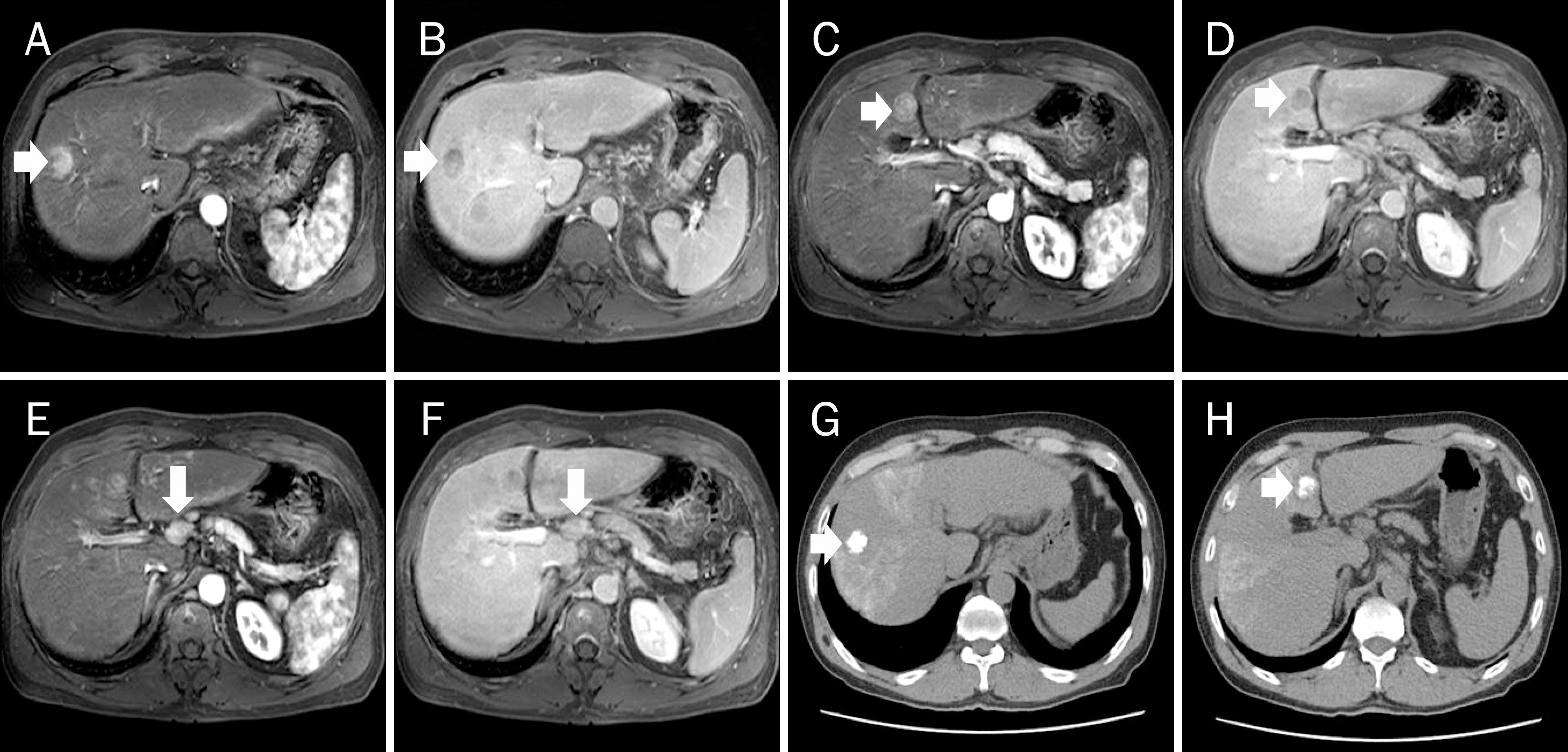
Fig. 1. Gadoxetic acid-enhanced magnetic resonance image shows a 2.3 cm sized hepatic nodule in S8 (white arrows), which is enhanced on arterial phase (A) and washed-out on equilibrium phase (B). Another 2.1 cm sized hepatic nodule in S4 (white arrows) is noted with same enhancement pattern on arterial (C) and equilibrium phase (D). An enlarged lymph node is also seen along the common hepatic artery, and it is enhanced on arterial phase (E) and iso-attenuated on equilibrium phase (F). After the first transarterial chemoembolization, the compact lipiodol uptake by both hepatic nodules is shown in pre-enhanced phase (G, H).
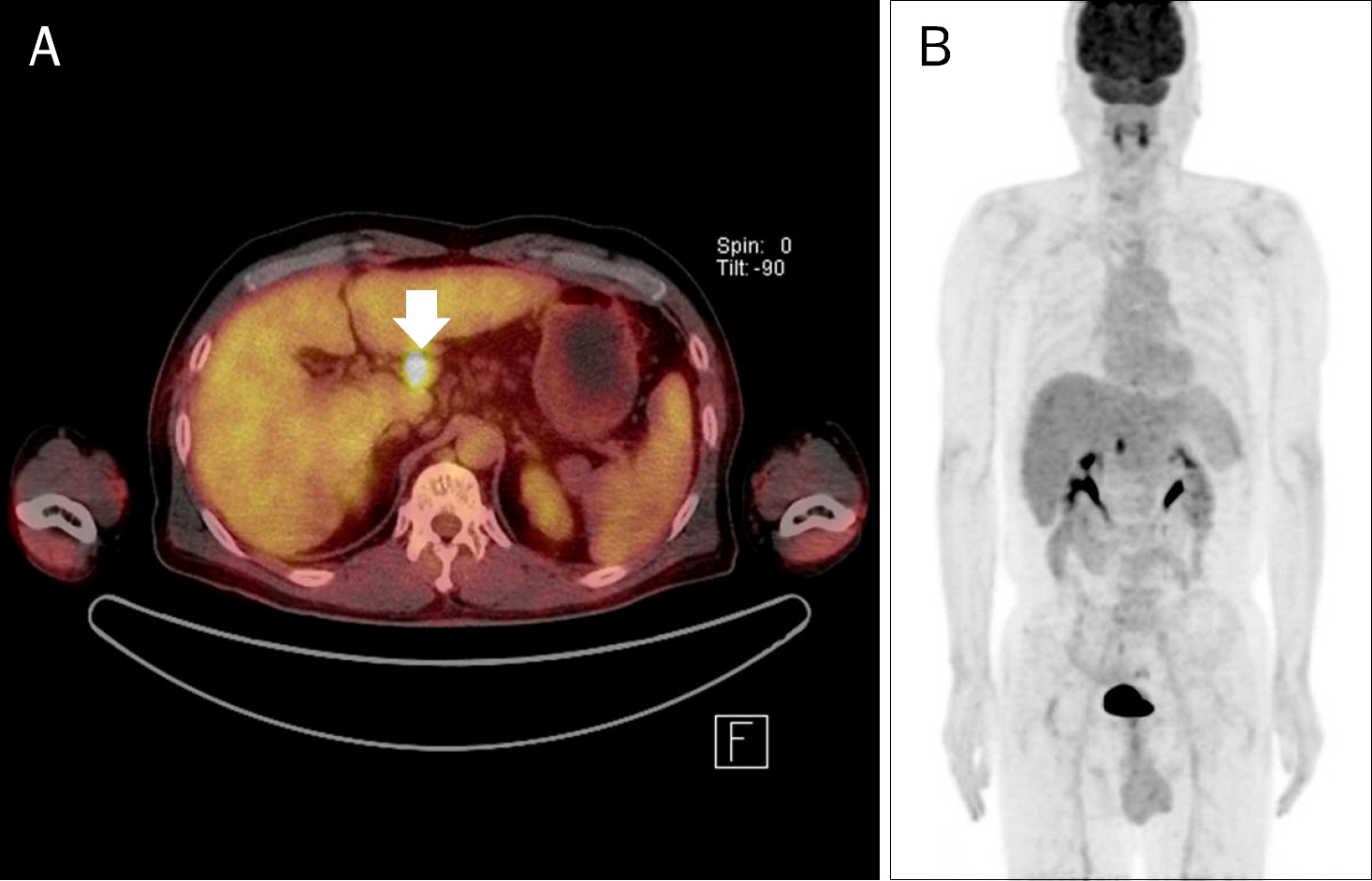
Fig. 2. (A) Axial and (B) maximum intensity projection image of 18 F-FDG PET-CT shows hyper-metabolic activity in the enlarged periportal lymph node (white arrow), the maximum standar-dized uptake value (SUVmax) of which is 6.8. The SUVmax of the hepatic nodules is 3.45, which is iso-metabolic compared to normal hepatic paren-chyma.
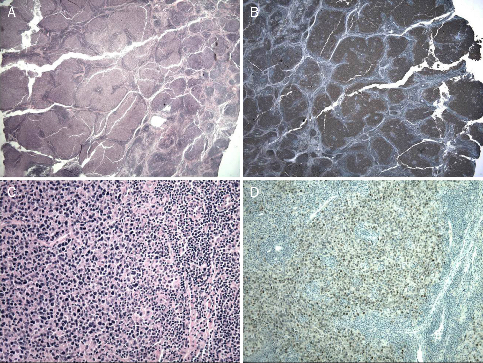
Fig. 3. Low-power photomicrograph of the periportal lymph node reveals characteristic nodular growth pattern in H&E (A; ×20) and CD20 immunohistochemical stain (B; ×20). At high-power microscopic view, each follicle is comprised predominantly of large cells resembling centroblasts (C; H&E, ×400). The neoplastic lymphocytes are positive for Bcl-6, which is expressed in germinal center cells (D; ×200).
Reference
-
References
1. Marcucci F, Mele A. Hepatitis viruses and non-Hodgkin lymphoma: epidemiology, mechanisms of tumorigenesis, and therapeutic opportunities. Blood. 2011; 117:1792–1798.
Article2. Ono T, Komatsu M, Masamune O. Primary lymphoma of the spleen with hepatocellular carcinoma. Intern Med. 1995; 34:261–264.
Article3. Suriawinata A, Ye MQ, Emre S, Strauchen J, Thung SN. Hepatocellular carcinoma and non-Hodgkin lymphoma in a patient with chronic hepatitis C and cirrhosis. Arch Pathol Lab Med. 2000; 124:1532–1534.
Article4. Shapira MY, Muszkat M, Braunstein I, Gotsman I. Co-occurrence of hepatocellular carcinoma and lymphoma in patients with hepatitis C virus cirrhosis. J Clin Gastroenterol. 2001; 32:368–369.
Article5. Himoto T, Miyauchi Y, Nomura K, et al. Coexistence of splenic non-Hodgkin's lymphoma with hepatocellular carcinoma in a patient with chronic hepatitis C. Dig Dis Sci. 2006; 51:70–76.
Article6. Ohtsubo K, Oku E, Imamura R, et al. Simultaneous hepatic relapse of non-Hodgkin's lymphoma and hepatocellular carcinoma in a patient with hepatitis C virus-related cirrhosis. Acta Haematol. 2006; 116:266–271.
Article7. Lin A, Kadam JS, Bodenheimer HC, Leonard J, Joyce MA, Lake-Bakaar G. Concomitant diffuse large B-cell lymphoma and hepatocellular carcinoma in chronic hepatitis C virus liver disease: a study of two cases. J Med Virol. 2008; 80:1350–1353.
Article8. Utsunomiya T, Okamoto M, Tsujita E, et al. Hepatocellular carcinoma infiltrated with non-Hodgkin's lymphoma: report of a case. Surg Today. 2009; 39:1010–1012.
Article9. Becker DJ, Sevilla DW, O'Connor O. Concurrent and apposed hepatocellular carcinoma and small lymphocytic lymphoma/chronic lymphocytic leukemia in a patient with hepatitis C virus. Acta Haematol. 2010; 123:77–80.
Article10. Ishikawa T, Shibuya K, Yasui K, Mitamura K, Ueda S. Expression of hepatitis C virus core protein associated with malignant lymphoma in transgenic mice. Comp Immunol Microbiol Infect Dis. 2003; 26:115–124.
Article
- Full Text Links
-
- Actions
-
Cited
- CITED
-
- Close
- Share
-
- Similar articles
-
- Relationships among Hepatitis C Virus, Hepatocellular Carcinoma, and Diffuse Large B Cell Lymphoma: A Case Report
- Case of Synchronous Primary Gastric Diffuse Large B-Cell Lymphoma and Hepatocellular Carcinoma
- A case of synchronous triple primary cancer of non-Hodgkin's lymphoma associated with hepatocellular carcinoma and early gastric cancer
- A Case of Non-Hodgkin's Lymphoma Associated with Hepatocellular Carcinoma
- A Case of Synchronous Gastric Non - Hodgkin's Lymphoma and Renal Cell Carcinoma
