Two-dimensional neovascular complexity is significantly higher in nontumor prostate tissue than in low-risk prostate cancer
- Affiliations
-
- 1Department of Urology, Humanitas Clinical and Research Hospital, Rozzano, Milan, Italy. gianluigi.taverna@humanitas.it
- 2Department of Inflammation and Immunology, Humanitas Clinical and Research Hospital, Rozzano, Milan, Italy.
- 3Department of Pathology, Humanitas Clinical and Research Hospital, Rozzano, Milan, Italy.
- KMID: 2160583
- DOI: http://doi.org/10.4111/kju.2015.56.6.435
Abstract
- PURPOSE
Prostate cancer is the most frequent cancer in men in Europe. A major focus in urology is the identification of new biomarkers with improved accuracy in patients with low-risk prostate cancer. Here, we evaluated two-dimensional neovascular complexity in prostate tumor and nontumor biopsy cores by use of a computer-aided image analysis system and assessed the correlations between the results and selected clinical and pathological parameters of prostate carcinoma.
MATERIALS AND METHODS
A total of 280 prostate biopsy sections from a homogeneous series of 70 patients with low-risk prostate cancer (Gleason score 3+3, prostate-specific antigen [PSA]<10 ng/mL, and clinical stage T1c) who underwent systematic biopsy sampling and subsequent radical prostatectomy were analyzed. For each biopsy, 2-microm sections were treated with CD34 antibodies and were digitized by using an image analysis system that automatically estimates the surface fractal dimension.
RESULTS
Our results showed that biopsy sections without cancer were significantly more vascularized than were tumors. No correlations were found between the vascular surface fractal dimension and patient's age, PSA and free-to-total PSA ratios, pathological stage, Gleason score, tumor volume, vascular invasion, capsular penetration, surgical margins, and biochemical recurrence.
CONCLUSIONS
The value of angiogenesis in prostate cancer is still controversial. Our findings suggest that low-risk prostate cancer tissues are less vascularized than are nontumor tissues. Further studies are necessary to understand whether angiogenesis is a hallmark of intermediate- and high-risk prostate cancer.
Keyword
MeSH Terms
-
Adult
Aged
Biopsy, Needle
Fractals
Humans
Image Processing, Computer-Assisted/methods
Kallikreins/blood
Male
Middle Aged
Neoplasm Grading
Neoplasm Staging
Neovascularization, Pathologic/*pathology
Prostate/*blood supply
Prostate-Specific Antigen/blood
Prostatectomy
Prostatic Neoplasms/*blood supply/pathology/surgery
Retrospective Studies
Kallikreins
Prostate-Specific Antigen
Figure
-
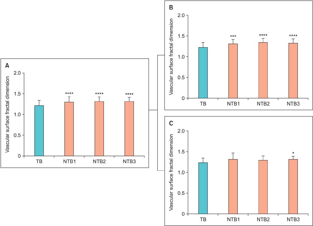
Fig. 1 (A) The surface fractal dimension, as a numerical index of the two-dimensional geometrical complexity of tumor vascular networks, in patients with prostate cancer. Statistically significant differences were found between the surface fractal dimension in tumor biopsy (TB) samples versus nontumoral biopsy (NTB) cores. The same difference is found when the population is divided in two groups according to the pathological Gleason grade 3+3 or 3+4 at the subsequent radical prostatectomy (B and C, respectively). *p<0.01. ***p<0.0001. ****p<0.00001.

Fig. 2 Multilevel-based procedure for estimating the two-dimensional vascular fractal dimension in tumor and nontumor biopsy cores. It has been recognized that two main issues influence results in the quantification of microvascularity in prostate tissue: (1) subjectivity in the diagnostic interpretation and (2) inadequate quantitative parameter. The present procedure automatically identified the microvascularity and estimated the surface fractal dimension as an index of the geometrical microvascular complexity.
Reference
-
1. Siegel R, Ma J, Zou Z, Jemal A. Cancer statistics, 2014. CA Cancer J Clin. 2014; 64:9–29.2. Stephan C, Ralla B, Jung K. Prostate-specific antigen and other serum and urine markers in prostate cancer. Biochim Biophys Acta. 2014; 1846:99–112.3. Eggener SE, Badani K, Barocas DA, Barrisford GW, Cheng JS, Chin AI, et al. Gleason 6 prostate cancer: translating biology into population health. J Urol. 2015; 04. 04. [Epub]. http://dx.doi.org/10.1016/j.juro.2015.01.126.4. Ahmed HU, Arya M, Freeman A, Emberton M. Do low-grade and low-volume prostate cancers bear the hallmarks of malignancy? Lancet Oncol. 2012; 13:e509–e517.5. Hanahan D, Weinberg RA. Hallmarks of cancer: the next generation. Cell. 2011; 144:646–674.6. Kulac I, Haffner MC, Yegnasubramanian S, Epstein JI, De Marzo AM. Should Gleason 6 be labeled as cancer? Curr Opin Urol. 2015; 25:238–245.7. Loeb S, Montorsi F, Catto JW. Future-proofing Gleason grading: what to call Gleason 6 prostate cancer? Eur Urol. 2015; 03. 10. [Epub]. http://dx.doi.org/10.1016/j.eururo.2015.02.038.8. van der Kwast TH, Roobol MJ. Prostate cancer: Is prostatectomy for Gleason score 6 a treatment failure? Nat Rev Urol. 2015; 12:10–11.9. Carmeliet P. Angiogenesis in health and disease. Nat Med. 2003; 9:653–660.10. Taverna G, Grizzi F, Colombo P, Graziotti P. Is angiogenesis a hallmark of prostate cancer? Front Oncol. 2013; 3:15.11. Grizzi F, Colombo P, Taverna G, Chiriva-Internati M, Cobos E, Graziotti P, et al. Geometry of human vascular system: is it an obstacle for quantifying antiangiogenic therapies? Appl Immunohistochem Mol Morphol. 2007; 15:134–139.12. Miyata Y, Mitsunari K, Asai A, Takehara K, Mochizuki Y, Sakai H. Pathological significance and prognostic role of microvessel density, evaluated using CD31, CD34, and CD105 in prostate cancer patients after radical prostatectomy with neoadjuvant therapy. Prostate. 2015; 75:84–91.13. Bostwick DG, Wheeler TM, Blute M, Barrett DM, MacLennan GT, Sebo TJ, et al. Optimized microvessel density analysis improves prediction of cancer stage from prostate needle biopsies. Urology. 1996; 48:47–57.14. Gettman MT, Bergstralh EJ, Blute M, Zincke H, Bostwick DG. Prediction of patient outcome in pathologic stage T2 adenocarcinoma of the prostate: lack of significance for microvessel density analysis. Urology. 1998; 51:79–85.15. Weidner N, Carroll PR, Flax J, Blumenfeld W, Folkman J. Tumor angiogenesis correlates with metastasis in invasive prostate carcinoma. Am J Pathol. 1993; 143:401–409.16. Di Ieva A, Grizzi F, Jelinek H, Pellionisz AJ, Losa GA. Fractals in the neurosciences, part i: general principles and basic neurosciences. Neuroscientist. 2013; 20:403–417.17. Waliszewski P, Wagenlehner F, Gattenlohner S, Weidner W. Fractal geometry in the objective grading of prostate carcinoma. Urologe A. 2014; 53:1186–1194.18. Grizzi F, Russo C, Colombo P, Franceschini B, Frezza EE, Cobos E, et al. Quantitative evaluation and modeling of two-dimensional neovascular network complexity: the surface fractal dimension. BMC Cancer. 2005; 5:14.19. Taverna G, Colombo P, Grizzi F, Franceschini B, Ceva-Grimaldi G, Seveso M, et al. Fractal analysis of two-dimensional vascularity in primary prostate cancer and surrounding nontumoral parenchyma. Pathol Res Pract. 2009; 205:438–444.20. Chambo RC, Tsuji FH, de Oliveira Lima F, Yamamoto HA, Nobrega de Jesus CM. What is the ideal core number for ultrasound-guided prostate biopsy? Korean J Urol. 2014; 55:725–731.21. Erbersdobler A, Isbarn H, Dix K, Steiner I, Schlomm T, Mirlacher M, et al. Prognostic value of microvessel density in prostate cancer: a tissue microarray study. World J Urol. 2010; 28:687–692.22. Taverna G, Grizzi F, Colombo P, Graziotti PP. Microvessel density estimate: friend or foe in the light of prostate vascular system complexity? World J Urol. 2010; 28:405–406.23. Hlatky L, Hahnfeldt P, Folkman J. Clinical application of antiangiogenic therapy: microvessel density, what it does and doesn't tell us. J Natl Cancer Inst. 2002; 94:883–893.24. Tretiakova M, Antic T, Binder D, Kocherginsky M, Liao C, Taxy JB, et al. Microvessel density is not increased in prostate cancer: digital imaging of routine sections and tissue microarrays. Hum Pathol. 2013; 44:495–502.25. Deering RE, Bigler SA, Brown M, Brawer MK. Microvascularity in benign prostatic hyperplasia. Prostate. 1995; 26:111–115.26. Barth PJ, Weingartner K, Kohler HH, Bittinger A. Assessment of the vascularization in prostatic carcinoma: a morphometric investigation. Hum Pathol. 1996; 27:1306–1310.27. Luczynska E, Gasinska A, Wilk W. Microvessel density and expression of vascular endothelial growth factor in clinically localized prostate cancer. Pol J Pathol. 2013; 64:33–38.28. Grizzi F, Chiriva-Internati M. Cancer: looking for simplicity and finding complexity. Cancer Cell Int. 2006; 6:4.29. Taverna G, Pedretti E, Di Caro G, Borroni EM, Marchesi F, Grizzi F. Inflammation and prostate cancer: friends or foe? Inflamm Res. 2015; 64:275–286.30. Lepor H, Donin NM. Gleason 6 prostate cancer: serious malignancy or toothless lion? Oncology (Williston Park). 2014; 28:16–22.
- Full Text Links
-
- Actions
-
Cited
- CITED
-
- Close
- Share
-
- Similar articles
-
- Milk Consumption and Prostate Cancer: A Systematic Review
- Prostate Cancer in Young Man
- Emerging Roles of Human Prostatic Acid Phosphatase
- A Continuous Increase in Prevalence of state Cancer in Korea and Its Causes
- Is a Decreased Serum Testosterone Level a Risk Factor for Prostate Cancer? A Cohort Study of Korean Men
