Obstet Gynecol Sci.
2016 Mar;59(2):144-147. 10.5468/ogs.2016.59.2.144.
A live birth after spontaneous complete chorioamniotic membrane separation associated with uterine scar
- Affiliations
-
- 1Department of Obstetrics and Gynecology, Ulsan University Hospital, University of Ulsan College of Medicine, Ulsan, Korea.
- 2Department of Obstetrics and Gynecology, Inha University Hospital, Inha University School of Medicine, Incheon, Korea. lenna@hanmail.net
- KMID: 2159010
- DOI: http://doi.org/10.5468/ogs.2016.59.2.144
Abstract
- Spontaneous complete chorioamniotic membrane separation (CMS) without invasive fetal procedure is extremely rare and associated with adverse perinatal outcomes. A woman with complete CMS which was detected at the 21 weeks' gestation. She did not take any fetal invasive procedures before the diagnosis. At 27 weeks' gestation, an emergency Caesarean section was performed because of fetal distress. The defect of the uterine muscle was detected on the fundus. The baby has grown well without any morbidity. This is the first reported case of complete CMS relative to uterine scar. And we suggest that the pregnancy can be maintained successfully if there is no fetal abnormality when complete CMS is detected on ultrasound.
Keyword
MeSH Terms
Figure
-
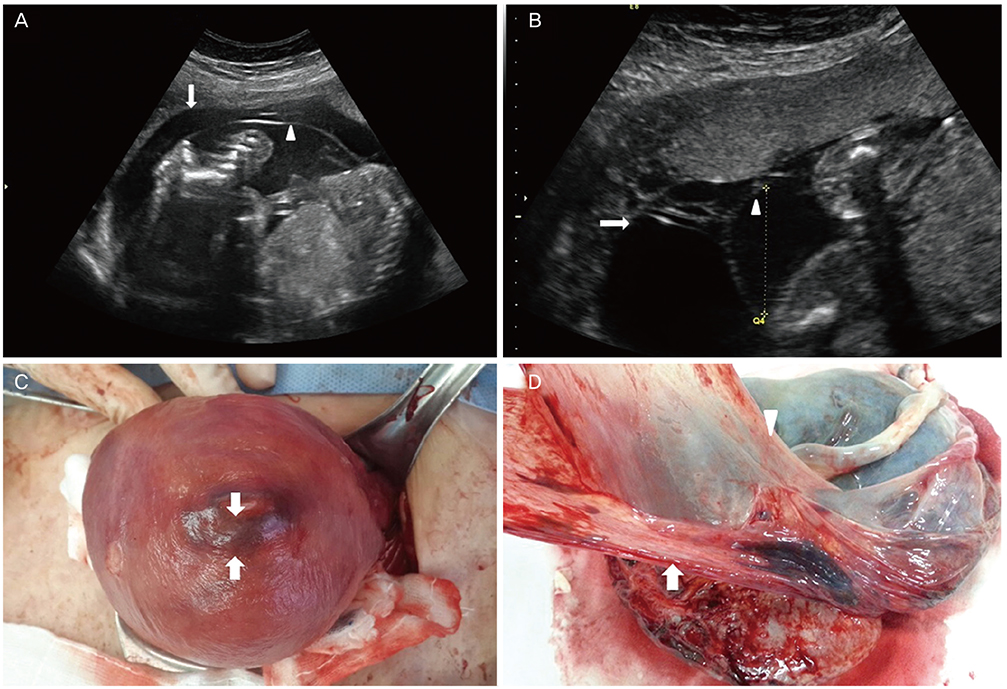
Fig. 1 Complete chorioamniotic membrane separation. (A) Ultrasonographic image at 21 weeks' gestation, showing the amnion (arrowhead) separated from the chorion (arrow) and fetus in amniotic cavity. (B) Ultrasonographic image at 21 weeks' gestation, showing normal-looking placenta and decreased amount of amniotic fluid volume in the amniotic cavity; chorion (arrow), amnion (arrowhead). (C) A localized mushy surface (arrow) measuring 1.5 cm in diameter was found on the fundus of the uterus, where was the defect of the uterine muscle. (D) Placenta reveals the amnion (arrowhead) separated from the chorion (arrow).
Reference
-
1. Kim YN, Jeong DH, Jeong SJ, Sung MS, Kang MS, Kim KT. Complete chorioamniotic membrane separation with fetal restrictive dermopathy in two consecutive pregnancies. Prenat Diagn. 2007; 27:352–355.2. Sydorak RM, Hirose S, Sandberg PL, Filly RA, Harrison MR, Farmer DL, et al. Chorioamniotic membrane separation following fetal surgery. J Perinatol. 2002; 22:407–410.3. Egawa M, Hayashi S, Yang L, Sakamoto N, Sago H. Chorioamniotic membrane separation after fetoscopic laser surgery for twin-twin transfusion syndrome. Prenat Diagn. 2013; 33:89–94.4. Abboud P, Mansour G, Zejli A, Gondry J. Chorioamniotic separation after 14 weeks' gestation associated with trisomy 21. Ultrasound Obstet Gynecol. 2003; 22:94–95.5. Bronshtein M, Zimmer EZ. Oligohydramnios with amniochorionic separation at 15-16 weeks' gestation. Prenat Diagn. 1995; 15:161–164.6. Lewi L, Hanssens M, Spitz B, Deprest J. Complete chorioamniotic membrane separation. Case report and review of the literature. Fetal Diagn Ther. 2004; 19:78–82.7. Bromley B, Shipp TD, Benacerraf BR. Amnion-chorion separation after 17 weeks' gestation. Obstet Gynecol. 1999; 94:1024–1026.8. Kasuga Y, Miyakoshi K, Ikenoue S, Kadohira I, Matsumoto T, Minegishi K, et al. Complete chorion-amnion separation presenting as a stuck fetus. Acta Obstet Gynecol Scand. 2013; 92:990–991.9. Ishikawa G, Satomi M, Inagawa-Ichikawa T, Abe T, Akira S, Takeshita T. A case report of complete chorioamniotic membrane separation. J Nippon Med Sch. 2011; 78:120–125.
- Full Text Links
-
- Actions
-
Cited
- CITED
-
- Close
- Share
-
- Similar articles
-
- Two cases of complete chorioamniotic membrane separation
- Chorioamniotic membrane separation caused by the seromucinous collection from a placental chorioangioma
- Spontaneous Separation of a Secondary Macular Epiretinal Membrane
- Spontaneous Separation of Epiretinal Membrane in Young Adult
- Reproductive Outcome of Women with Recurrent Abortions or Infertility Following Treatment by Operative Hysteroscopy for an Intrauterine Septum
